Human skeleton of the thoracic region. What is the human skeleton made of
The skeleton is a frame made of bones on which our body rests. It gives the body shape and stability, and also protects the internal organs.
What is a Skeleton?
The body owes its hardness, shape and strength to the skeleton. The skeleton consists of three main parts. The spine and ribs form axial skeleton, with which the skull and bones of the limbs are docked. The structure of 206 hard, strong bones forms the internal frame of the body and protects the delicate internal organs - the brain, heart, lungs, etc. The bones are connected to each other by joints, and the muscles that set the body in motion lean to the bones.
Bone strength
The size, shape and strength of a bone depend on which part of the body it supports and which muscles are attached to it. The hardness of bones is due to the presence of crystals of calcium salts and phosphates. But the bones have some flexibility, because they contain elastic protein tissues, and when loaded, they spring slightly, and do not break.
What are bones made of?
The bones are painted pale yellow, have their own blood vessels and nerves. Bone is a combination of living cells with mineral formations. Bone cells, the so-called osteocytes, produce miniature bone plates that form osteons (the Haversian system). Appearance most bones - this is a strong substance in which osteons are located very tightly. Under it is a loose spongy substance.
Bone marrow
The tubular bones contain a jelly-like Bone marrow producing millions of blood cells every second. In a newborn, red bone marrow is present in all bones; in an adult, it is preserved only in the sternum, spine, ribs, skull and pelvic bones.
Human skeleton
The cranium consists of 8 bones fastened together, the face consists of 14. Inside each ear are 3 tiny bones of the skeleton. The spine is made up of 26 vertebrae. 12 pairs of ribs connect to the sternum at the front chest. There are 32 bones in each hand, 8 of which are in the wrist. There are 31 bones in each leg, 7 of which are in the tarsus.
Skeleton plays the role of supporting the human body. In addition, it has a protective function for many important parts of the body and organs. So, the cranium protects the brain, the chest protects the heart and lungs from mechanical influences (shocks, pressure, etc.).
The human skeleton is made up of bones, cartilage and ligaments.. Bones are mainly formed bone tissue, the structure of which was described earlier. Bones are distinguished by great strength, depending on the fact that their substance combines the hardness of inorganic salts (mainly calcium) with elastic properties. organic compounds. Most bones have dense and spongy parts. In the first, the bone tissue forms a solid solid mass or layers without visible cavities. In the spongy parts of the bones, bone tissue forms complex systems of mutually intersecting plates and crossbars, between which there are small cavities filled with red bone marrow, in which hematopoiesis.
There are three types of bones in the human skeleton:
long bones. Their middle part has a tubular structure. The walls of the tube are formed by dense bone tissue, and the median cavity is filled with yellow bone marrow, which is rich in fat. Both ends of the long bones are covered on the outside with a strong layer of dense bone tissue, and inside they are built of spongy bone substance. The correct vaulted arrangement of the crossbars of the bone substance ensures greater strength of the heads of long bones with their relatively low weight. The long bones include the humerus, forearm bones, femurs, and lower leg bones.
flat bones have the character of bone plates. On both sides they are formed by layers of dense bone tissue, and in the middle part they usually have a spongy structure. These are the bones of the cranium, shoulder blades, sternum.
short bones- small bones, the length of which is only slightly greater than the width or approximately equal to it. Outside, they consist of a layer of dense bone tissue, and inside they are filled with spongy bone mass.
Bones are covered with a thin layer of connective tissue periosteum. Usually, in embryos, bones are laid in the form of a soft rudiment of fibrous connective tissue, which is later replaced by cartilage, and the cartilage then ossifies.
As a person grows, the length and thickness of bones increase. Their length increases due to the fact that a still growing person has at both ends tubular bones there are layers of cartilage. Their cells multiply towards the ends of the bone, and on the opposite side of the layer, the cartilage is replaced by bone, as a result of which the length of the bone increases. The thickness of tubular bones increases due to new layers of bone tissue deposited by the periosteum.
Skeletal cartilage formed by special cartilage tissue. Its oval cells lie in capsules among a solid elastic intermediate substance. Sometimes this substance seems amorphous, translucent (the so-called hyaline cartilage), in other cases a network of the thinnest fibers (fibrous cartilage) is visible in it. Articular surfaces of bones are covered with cartilage, strips of cartilage often connect the ends of bones, layers of cartilage are located between the vertebrae.
The human skeleton is divided on: the skeleton of the body, the skeleton of the limbs and their belts and the skeleton of the head (skull). The skeleton of the body consists of the spine and chest.
human spine consists of 7 cervical, 12 thoracic, 5 lumbar, 5 sacral and 4-5 coccygeal vertebrae. sacral vertebrae fuse into a single bone sacrum. An individual vertebra consists of a massive body, a bony arch, and several processes. Holes between the bodies and arches of individual vertebrae, located one above the other, form the spinal canal, inside which is spinal cord. There are layers of cartilage between the vertebrae.
Thoracic vertebrae, ribs and sternum form the chest. The ribs (there are 12 pairs in humans) are flat, arcuately curved long bones, which are movably articulated behind their heads with the thoracic vertebrae, and in front (except for the two lower pairs) are connected with flexible chryaforms, lying on the back surface of the chest. On the upper outer corner they have fossae, where the heads of the humerus enter. The clavicles are connected at one end to the upper end of the sternum, and at the other - to the shoulder blades. Skeleton upper limb comprises humerus, two bones of the forearm (ulna and radius), a number of small bones of the wrist of five parallel long bones of the metacarpus and phalanges of five fingers.
Belt of the lower extremities formed by a pair of massive pelvic bones, which are fused with the sacrum, forming a pelvic ring. On the sides of the pelvic bones there are pits for articulation with the femurs. The skeleton of the lower extremities is composed of large femur bones, lower leg bones (tibia and tibia), a number of tarsal bones, long bones of the metatarsus and phalanges of the fingers.
The bones of the limbs are movably connected joints. The articulating surfaces of the bones are covered with a layer of smooth cartilage. The joint is surrounded by a strong articular bag. In addition, the bones that form the joint are connected by elastic but strong ligaments. The joint capsule contains joint fluid inside, which reduces the friction of the articular surfaces of the bones.
The skeleton of the head is called the skull; it is subdivided into brain and facial sections. brain department- cranium - contains the brain and protects it from shock, pressure and other influences. It is formed by a series of flat bones fixed to each other. In front lies a large unpaired frontal bone. Above, behind the frontal bone, are the paired parietal bones. The posterior lower part of the skull is formed occipital bone with a large occipital foramen, in which the brain and spinal cord are connected. On the sides of the skull are paired temporal bones with openings for the ear canal. The floor of the skull is formed by a series of bones with openings for the passage of nerves and blood vessels.
The facial section of the skull consists of the lower and upper jaws, zygomatic bones, nasal bones, etc. All these bones, except for the mandibular, are fixedly connected to each other. Lower jaw articulates flexibly with the temporal bones.
With various mechanical influences (impact, strong pressure, etc.) or awkward movement, joint sprains, dislocation or fracture of bones can occur. When the joint is stretched, the wall of the articular bag or ligament that connects the bones is damaged. The joint swells up sharp pain joint movements become difficult. With a dislocation in the joint, the head of one bone comes out of the glenoid fossa of the other.
When the bones of the limbs are fractured, there is a sharp pain, the damaged area swells, sometimes the limbs are bent or take an unnatural position. In all cases of damage to bones and joints, an urgent need to call a doctor. But before his arrival, first aid should be provided to the victim. In case of sprain and dislocation of the joint, a cold compress is placed on it and the limb is tightly bandaged. When a bone of a limb is fractured, it must be covered with something soft, smooth boards or a special splint should be applied to it and bandaged to the limb with a wide bandage, towels, strips of fabric.
Supports as well as protection. Movement function carried out with the help of bones articulated by joints and contraction of muscles attached to them. Support function consists in attaching soft tissues and organs to various bones of the skeleton. Protection function It is expressed in the formation of cavities in the bones, in which the vital organs are located. So, the chest protects the heart and lungs from mechanical influences, the cranium - the brain, etc. Bones are also a source of minerals. They contain red bone marrow involved in hematopoiesis.
There are over 200 bones in the human skeleton. They are formed by bone tissue, which includes organic substances (ossein, osseomucoid, etc.) and inorganic compounds (mainly calcium carbonate and phosphate). Organic substances give the bones flexibility and elasticity, inorganic - hardness. The share of organic substances in bone mass is about 30%, the remaining 70% are inorganic compounds. With age, the proportion of inorganic substances increases, and organic decreases, which makes the bones more fragile and difficult to fight after fractures.
The structure of the bone. On a longitudinal cut of a tubular bone (Fig. 12.4), two types of bone substance are well distinguished: outside - dense compact and inside- spongy. Both types of substance consist of loosely located bone cells and the intercellular substance secreted by them with immersed in it with protein fibers. Together, these elements form bone plates, and they, in turn, are larger bone bars, or beams. In the spongy substance, the crossbars are located loosely, forming between themselves cells like a sponge. If the crossbars fit snugly together in the form of concentric circles around the channels in which the nerves and blood vessels that feed the bone pass, then a compact bone substance is formed. The compact substance, being outside, gives strength to the bone, and the spongy substance reduces the mass of the bone. The ratio of dense and compact bone substance is different for different bones and depends on their shape, function and location.
Outside, the bone, with the exception of the articular surfaces, is covered periosteum. It is a dense connective tissue sheath, which is fused with the bone through collagen fibers. The periosteum contains many blood vessels that penetrate the thickness of the bone and feed it. In the inner layer of the periosteum there are cells (osteoblasts) capable of forming new bone cells. Therefore, the periosteum ensures the growth of bones in thickness, as well as the healing of bone fractures.
Rice. 12.4. Scheme of the structure of the tubular bone: 1 -spongy substance; 2 - compact substance; 3 - medullary cavity; four - Bone marrow ; 5 - periosteum; 6-blood vessel; 7-nutrient hole.
Bone contains two types of marrow. The cells between the crossbars of the cancellous bone are filled red bone marrow. It has many blood vessels that feed the bone from the inside, as well as hematopoietic cells. The cavity of tubular bones contains yellow marrow, represented mainly by fat cells, giving it a yellow color.
Bone shape. According to the shape of the bones of the skeleton are divided into tubular, flat and mixed.
tubular bones divided into long and short. Long tubular bones that form the basis of the limbs act as levers set in motion by muscles (bones of the shoulder, forearm, thigh, lower leg). These bones have thickened ends - heads, or epiphyses, and a hollow (in the form of a tube) middle part - the body, or diaphysis, the walls of which are formed by a compact substance. Being light, such bones are able to provide great resistance to compression and stretching. During the period of bone growth, cartilaginous layers are located between the body and heads. Cartilage cells divide towards the ends of the bone, and on the opposite side of the layer, the cartilage is replaced by bone, resulting in an increase in the length of the bone. Complete ossification of the human skeleton occurs by the age of 20-25. Short tubular bones are located in places where high mobility is combined with resistance to compressive forces (tarsal bones, wrists).
flat bones form protective cavities for internal organs (skull bones, pelvic bones, ribs, shoulder blades, etc.).
To mixed belong to bones formed from several parts with different structures and functions (temporal, sphenoid bones).
Connection of bones. There are three types of bone connections: fixed, semi-movable and mobile, or joint (Fig. 12.5).
Fixed connections are carried out by bone fusion (sacral vertebrae), as well as sutures (skull bones). They provide connection reliability and the ability to withstand heavy loads.
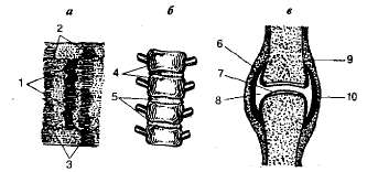
Figure 12.5. Fixed (a), semi-movable (b) and movable (c) connection of bones: 1-3 - sutures between the parietal, frontal and parietal and between the occipital and parietal bones, respectively; four - vertebrae; 5 - cartilaginous layers between the vertebrae; 6.7-articular surfaces; eight - articular cavity; 9 - periosteum; ten - articular bag.
semi-movable are called connections of bones with the help of cartilage (connection of the vertebrae in the spine, ribs with the sternum).
Joint - the most common and complex form of bone connection, providing a movable connection. Joints, regardless of differences in mobility, consist of three essential elements: articular surfaces, articular capsule and articular cavity (see Fig. 12.5). Articular surfaces articulating bones are ideally fitted in shape and fit snugly together. They are covered with special (hyaline) cartilage. Their smooth surface facilitates movement in the joint, and the elasticity of the cartilage softens the jolts and jolts experienced by the joint. connective tissue joint bag stretched between the articulating ends of the bones and attached to the edge of the articular surfaces, where it passes into the periosteum. In most joints, the bag is reinforced on the outside with ligaments. Articular cavity sealed and surrounded by articular cartilage and articular bag. It contains a small amount of a viscous fluid that lubricates the articular cartilage, which reduces friction in the joints during movement. Due to the negative pressure in the articular cavity, the surfaces of the articulating bones closely adjoin each other.
According to the shape of the articular surfaces, there are four types of joints: flat(between the bones of the wrist and metacarpus), cylindrical(joint between ulna and radius) elliptical(the articulation between the bones of the forearm and hand) and spherical(shoulder and hip joints). The smallest mobility is provided by flat joints, the greatest - by spherical ones.
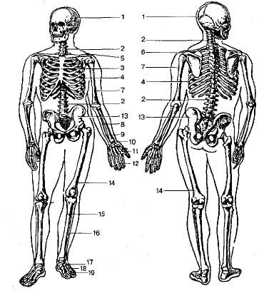
Figure 12.6. Human skeleton (front and back): 1 - skull; 2-spine; 3 - sternum; 4 - chest; 5 - collarbone; 6 - scapula; 7 - brachial bone; eight -elbow bone; 9 -radius; 10 - bones of the wrist; eleven - metacarpus bones; 12 - phalanges of fingers; 13-pelvic bone; fourteen - femur; fifteen - tibia; 16 - small tibia; 17-tarsal bones; eighteen- bones of the metatarsus.
The structure of the human skeleton and its features. Three sections are distinguished in the skeleton: the skeleton of the trunk, upper and lower extremities and the head - the skull (Fig. 12.6).
Torso skeleton consists of the vertebral column and chest. The spinal column is the support of the body. It is formed by 33-34 vertebrae and has 5 sections: cervical - 7 vertebrae, thoracic - 12, lumbar - 5, sacral - 5 and coccygeal - 4-5 vertebrae.Each vertebra is from the body and arcs. Seven processes depart from the vertebra: two transverse, unpaired spinous and two upper and lower articular processes. With the help of the latter, the vertebrae are articulated with each other. There is a vertebral foramen between the body and the vertebral arch. A collection of vertebral foramina located one above the other forms spinal canal, in which the spinal cord is located. The size of the vertebral bodies increases from the cervical to the lumbar due to the increasing load on the lower vertebrae. The vertebral bodies are interconnected by cartilaginous intervertebral discs, which ensure its mobility and flexibility. The sacral and coccygeal vertebrae are fused together and form the sacral and coccygeal bones.
In connection with the upright posture of a person, his spine has four bending. in the neck and lumbar regions the curves are convex forward (lordosis), in the thoracic and sacral - convex backwards (kyphosis). Thanks to the S-shaped spine, shocks are softened when walking, jumping and running, it is easier to maintain body balance and the volume of the cavity of the chest and pelvis increases.
Thoracic vertebrae, 12 pairs of ribs and sternum together form chest. Flat, arcuately curved ribs are articulated with the transverse processes of the bodies of the thoracic vertebrae. Upper ribs - 7 pairs - directly connected with chest a flat bone lying in the midline of the chest. The 8th-10th pairs of ribs located below them are connected to each other by cartilage and attached to the 7th pair of ribs. The 11th and 12th pairs of ribs do not connect to the sternum and are placed freely in soft tissues. The chest protects the heart, lungs, trachea, esophagus and large blood vessels located in it. Due to the rhythmic raising and lowering of the ribs, the volume of the chest changes. In connection with the upright posture of a person, its shape is flat and wide.
Upper limb skeleton includes the shoulder girdle and the skeleton of the free upper limbs (hands). The shoulder girdle is represented by two paired bones - shoulder blades and clavicle. The shoulder blade is a flat triangular bone, the outer angle of which forms the articular cavity for articulation with the head of the humerus. The clavicles are connected at one end to the sternum, and at the other to the scapula, thanks to which the human hand is able to perform various movements in three planes. The skeleton of the free upper limb is formed humerus, forearm, consisting of the ulna and radius bones, as well as brush. Eight short tubular bones are distinguished in the hand wrist, arranged in two rows of four bones, five long bones metacarpus, each of which has three phalanx fingers (except thumb with two phalanges).
Skeleton of the lower extremities consists of a pelvic girdle and free lower limbs (legs). Pelvic girdle formed by a pair of massive pelvic bones, which are fused to the sacrum at the back, and connected to each other in front with the help of cartilage (pubic fusion). In a growing organism, the pelvic bone consists of three bones connected by cartilage: iliac, ischial and pubic. At the site of their fusion there is a recess - acetabulum, serving to connect with the head of the femur. Due to upright posture, the human pelvis is wide and cup-shaped. The female pelvis is wider and shorter in shape, while the male pelvis is longer and narrower.
Skeleton Free lower limb comprises femur, leg bones(tibia and tibia) and foot bones(seven bones tarsus, five metatarsus and phalanx fingers). The foot has an arch formed by a support on the protrusion of the calcaneus and the anterior part of the metacarpal bones. The arched foot softens the shocks of the body when walking.
Head skeleton (skull) consists of two departments: cerebral and facial. brain department four times the volume of the front (in monkeys they are equal). The brain skull is formed by two paired bones (parietal and temporal) and four unpaired (frontal, occipital, ethmoid and sphenoid). Part facial department skull, which forms the bony skeleton of the face, includes three unpaired bones ( lower jaw, vomer and hyoid) and six paired bones (maxillary, palatine, zygomatic, nasal, lacrimal and inferior nasal conchas). In the bones of the upper and lower jaws, there are 16 cells for the necks and roots of the teeth. All bones of the skull, with the exception of the lower jaw, are fixedly connected. The lower jaw is connected by a joint with the processes of the temporal bones (Fig. 12.7).
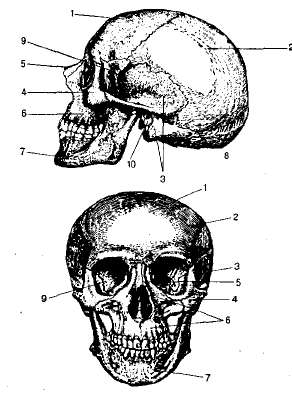
Rice 12.7. Skull front and side: 1 - frontal bone; 2 - parietal bone; 3 - temporal bone; four - cheekbone; 5 - nasal bone; b - upper jaw; 7 - lower jaw; eight - occipital bone; 9 - main bone; ten - external auditory canal.