How the circulatory and respiratory systems of reptiles become more complex. Biology at the Lyceum
Peculiarities internal structure and life activities of reptiles
Nutrition and digestion. Digestive systems reptiles and amphibians are similar in all main divisions. These are the mouth, pharynx, stomach, intestines. Food gets wet in the mouth saliva , which is typical of terrestrial animals. In the stomach under the influence gastric juice Protein foods are digested in an acidic environment. The ducts of the gallbladder, liver and pancreas open into the intestine. Here food digestion is completed and nutrients are absorbed into the blood.
Lizards eat mainly insects and worms. Snakes hunt voles and mice. Some snakes have special sensory pits on the front of their heads - thermolocators , capable of perceiving heat (infrared radiation) coming from a warm-blooded animal.
Venomous snakes kill their prey with venom flowing down the poisonous teeth
from poisonous glands
located in the walls of the oral cavity.
Respiratory system. Due to the emergence cervical spine The lizard's respiratory tract lengthens, through which air flows from the mouth to the lungs. Air is drawn in through the nostrils, into the mouth, then into larynx , then into a long tube - trachea ; the trachea is divided into two even narrower tubes - bronchi , going to the lungs. The lungs of reptiles are more complex than those of amphibians: inside the lung cavity there are many folds where they branch many times blood vessels. This increases the surface of their contact with air, enhancing gas exchange.
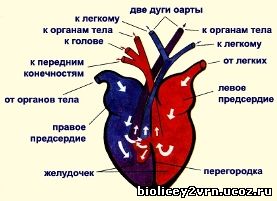 Circulatory system. Heart three-chamber
, with an incomplete septum in the ventricle. Three large vessels emerge from it: the left and right aortic arches and pulmonary artery. The two arches of the aorta, bypassing the heart, merge into one common vessel - the dorsal aorta.
Circulatory system. Heart three-chamber
, with an incomplete septum in the ventricle. Three large vessels emerge from it: the left and right aortic arches and pulmonary artery. The two arches of the aorta, bypassing the heart, merge into one common vessel - the dorsal aorta.
Mixed blood flows through the body (like in amphibians), which affects fickle temperature body , depending on the ambient temperature.
The pulmonary arteries carry venous blood from the heart to the lungs for oxygenation. The pulmonary veins carry arterial blood into the left atrium. In the ventricle, the blood is partially mixed, the most oxygen-rich blood goes to the head, mixed - to all organs of the body, saturated with carbon dioxide - to the lungs.
Excretory system. In reptiles, the excretory system is the same as in all terrestrial vertebrates. In the excretory organs - the kidneys - the mechanism for returning water to the body is strengthened: it is absorbed by the renal tubules. The end product of metabolism in reptiles is excreted not in the form of liquid urine (as in amphibians), but as uric acid in a pasty state into the cloaca, and then out. Eliminating mushy uric acid from the body does not require as much fluid as liquid urine.
 Nervous system. In reptiles, all parts of the brain become more complex and enlarged compared to the brain of amphibians. This is manifested in more complex and diverse behavior of reptiles. They form conditioned reflexes faster than fish and amphibians. The forebrain and cerebellum are especially enlarged, medulla oblongata forms a bend characteristic of all higher vertebrates. In addition to vision and smell, reptiles have a well-developed sense of touch.
Sense organs. The lizard's nostrils and tongue are involved in the perception of smells. The eyes are protected by the outer eyelids and the nictitating membrane. The secretions of the lacrimal glands protect the eyes from drying out. There is a special formation in the eye - lens , which changes shape under the action of a special muscle and ensures image clarity. The hearing organs consist of the inner and middle ear. Auditory ossicle - stapes – transmits sound vibrations from the eardrum to inner ear. In inner ear There are receptors that perceive sound.
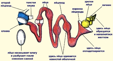
Reproduction and development. Reptiles are dioecious. In males, the reproductive organs are paired testes and vas deferens. The reproductive organs of females are two ovaries and oviducts. Oocytes develop in them. Fertilization in reptiles internal . It occurs when the cloaca of a male and female come together. The embryo developing in the fertilized egg, moving along the oviduct, is covered with egg and embryonic membranes. They provide the embryo with water, protect it from drying out, shaking, and participate in respiration and the release of metabolic products. The presence of the embryonic and protective membranes ensures the normal development of the embryo even in the absence of water. Development is direct, without the formation of larvae.
Reptiles lay eggs on the ground or in specially prepared holes. Some reptiles protect their clutches (for example, crocodiles); others, having laid eggs, leave them (for example, turtles). The eggs hatch into fully developed animals similar to their parents. Sometimes the cubs are carried in the mother's body. In these cases, live birth occurs, for example in vipers and at viviparous lizard
.
Annual life cycle. Reptiles are widespread around the globe and are found in different climatic zones. However, being cold-blooded animals with an unstable body temperature, they need external heating from the sun. Therefore, these animals are most numerous in the tropical and subtropical zones of the globe. In the changing seasons, when warm summer is replaced by cold autumn and winter, reptiles, with the onset of unfavorable conditions, go into shelters: holes, caves, under tree roots, under rural houses and forest huts. There the animals fall into stupor - hibernation . In the spring, when the air and soil surface warm up well, reptiles come to the surface and begin an active lifestyle.
Males and females in spring spinning lizards They wake up after hibernation and begin to mate. During the mating season, adult lizards split into pairs and settle together in one burrow, in the vicinity of which they hunt together and bask in the sun. At the end of May - beginning of June, the female lays from 6 to 16 eggs, burying them in a shallow hole or leaving them in the depths of the hole. Offspring usually appear at the end of July.
Target: reveal features external structure reptiles associated with a terrestrial way of life, show features of complexity in their organization compared to amphibians; improve the ability to recognize the studied species of reptiles in tables, drawings, compare them with each other and amphibians, compose general characteristics class.
Equipment: drawings “Class of reptiles”, macro-preparation “Viper”, multimedia, Korchagin’s textbooks, stories.
Lesson progress
I. Organizational moment (writing down homework).
II. Learning new material.
1) Activation of cognitive activity.
Previously, zoologists did not see significant differences between these animals and amphibians and always classified them as one group, uniting them under different names. Most often they appeared under the name “reptiles”. There have been attempts to divide reptiles into two separate classes - “holo-skinned”, or naked reptiles, and “hard-skinned” reptiles. Karl Lineaeus, characterizing his “class of reptiles”: “cold blood, lung breathing,” thereby noted their closeness in biological terms.
? Who are they? Why were they combined into one group “reptiles” along with amphibians?
Answer: these are reptiles.
2) Student message.
They could be called "vermin" for their appearance, which disgusted many. Let us remember amphibians, “disgusting, terrible, nasty and harmful.” The blood is cold - this means that all the “reptiles” are active only in the warm season, and the words “pulmonary breathing” characterize animals that lead a terrestrial existence.
Unusual behavior, strange appearance, often poisonousness - all this, as we know, made toads, frogs and salamanders attributes of witchcraft. Therefore, the attitude towards them was, at best, disgusting. The attitude towards reptiles was no better, if not worse.
3) The teacher writes a word on the board "Reptiles". [Slide 2]
Write down the associations that arise when you first perceive this term.
? What are reptiles? Why did they get this name?
Answer students.
I'm introducing a new term reptiles(crawling). [Slide 3]
4) Let's look at the appearance of reptiles.
(work in pairs)
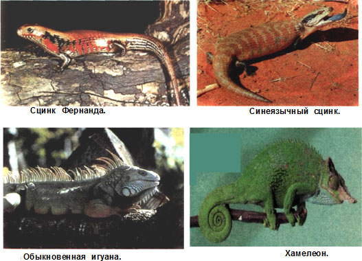
Working with Images (project a picture with images onto the screen[Slide 4] ) , which depicts a sharp-faced viper, a frog, a marsh turtle, a crested newt, a common toad, an alligator or rare and endangered species of amphibians and reptiles.
? Do all of the animals depicted belong to the class of reptiles?
Answers: (As students answer, numbers related to the class reptiles are written on the board). Let's check the correctness of your answers at the end of the lesson.
5) So, reptiles or reptiles. We need to find out what is behind these words? What characterizes these animals? What signs can characterize them? (Or students can formulate the objectives of the lesson themselves with the help of the teacher).
We write down the topic of the lesson [Slide 5].
External structure of reptiles.
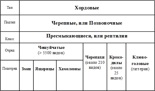
6) Currently, there are about 6,000 species of reptiles on Earth, that is, almost three times more than modern amphibians. Living reptiles are divided into 4 orders: scaly reptiles, turtles, crocodiles and beaked reptiles.
(A table is projected on the screen[Slide 6] )
7) ? In what climatic zones and under what conditions do these animals live?
A picture of the habitat and the animal is projected onto the screen [Slides 7-11].
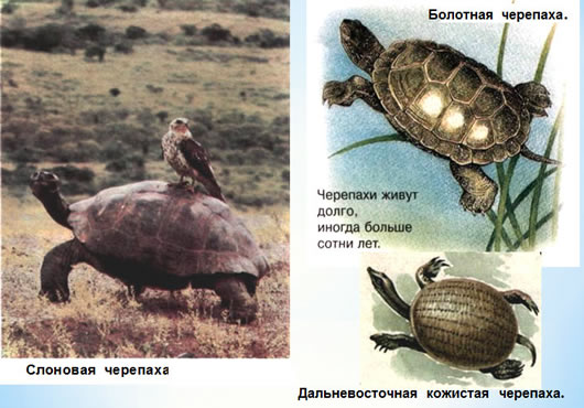
Turtles – from southern latitudes. Usually in open spaces, steppes, savannas, deserts. There are very few species in damp and wooded areas, on mountain slopes covered with bushes and open forests. In the water.
. Lizards – on all continents except Antarctica. There are more species in the tropics and subtropics, fewer species in temperate climates, and in the Arctic Circle there is only one species - the viviparous lizard.
They live in deserts, tropical forests, subalpine meadows, in gorges and mountains, in the seas (in the Galapagos - marine iguanas), in trees (chameleons).
Crocodiles – in the water.

(Message using a geographic map). Snakes – in almost all living spaces. There are many of them in the tropics of Asia, Africa, South America, Australia. In the same environments as lizards.

? Which sign, reflecting the habitat of these animals, can be formulated?
Answers students.
From the students' answers, the first sign is formulated to be written in the notebook. The wording of the first sign is projected on the screen [Slide 12], and students write it down in their notebooks.
1. Reptiles, as a rule, are land animals, i.e. lead a terrestrial lifestyle.
8) ? How do these animals move in a terrestrial environment? ?The teacher projects diagrams of the location of the limbs on the screen (1 – amphibians; 2 – reptiles)[Slide 13].
? What is shown in the pictures ?
Answers students.
We identify the features of the movement of reptiles and formulate next sign. Students write in their notebooks [Slide 14].
2. The short limbs of reptiles, located on the sides of the body, do not lift the body high above the ground, and it drags along the ground.
9) Physical education minute.
10) We find out how reptiles solve problems associated with life on land. The most main problem Drying is known to be a problem. To protect the body from excessive evaporation, reptiles covered themselves with scales. Their skin is thicker and harder, rough to the touch.
Let's test this assumption using the story "Leather Armor". The students have sheets of paper with the story on their desks and they read them.
? What features does the skin have? Why?
Answers students.
Let us formulate the third sign. Students write it down. [Slide 15]
3. The skin of reptiles is dry and has practically no glands; covered with horny scales.
11) Why is such skin necessary? What other significance does such skin have in the life of reptiles?
We look at two pictures [Slide 16], students express their opinions.
The teacher summarizes and complements the answers.
Scales and scutes are also important in the movement of reptiles, so their structure largely depends on the methods of movement and the characteristics of the animal’s habitat. Snakes and many lizards, without (or not using) limbs, rest on the ground with the ventral side of the body, and therefore they have developed wide and strong abdominal scutes [Slide 16]. They protect internal organs, but most importantly, with their help, the wriggling animal pushes off from uneven soil and crawls forward (a snake cannot crawl on a completely smooth surface). In those species that do not literally have to crawl on their bellies, such scutes are not needed and they are not developed. For example, sea snakes, which swim their entire lives in the thickness of ocean waters, and blind snakes, living in the soil, have their entire body covered with uniform small scales. If lizards or snakes have to constantly make their way through any obstacles - thickets of grass, intertwined branches, heaps of fallen leaves - then their skin is usually completely smooth, the scales fit tightly to one another. The main thing is not to get caught on anything, not to slow down the rapid slide. Taken in hand, such a reptile just flows through your fingers, slips out like soap. Such “slipperiness” is absolutely useless for tree snakes - after all, they cannot cling to branches with their paws and would simply fall to the bottom if the roughness of their skin did not help them stay on the tree.
Thus, the structural features of the skin are closely related to the method of movement of the reptile.
If the skin does not stretch, how does animal growth occur? What is shown in these pictures? What is this process? [Slide 17]
Answers students.
We formulate the fourth feature of the class [Slide 18].
4. D Reptiles are characterized by molting; growth of the body occurs during molting.
12) Another sign that we haven’t talked about in class yet. Students work from the textbook, chapter “Features of the external structure.”
Find this sign in the text. What significance does it have in the lives of animals?
Answers students.
Checking the fifth sign [Slide 19]:
5. The eyes of reptiles have eyelids that protect the eyes from damage.
13) Let's summarize. We summarize all the signs that we studied during the lesson. One of the students reads out all the signs. [Slide 20]
14) We check the correctness of the answers of the selected images at the beginning of the lesson. [Slide 21]
15) Reflection. [Slide 22]
Appearance
The lizard's body is clearly divided into a head, neck, torso, tail and paired limbs - front and rear.
:
1 - external nostrils, 2 - eyes, 3 - external ear opening, 4 - claws,
5 - horny scales, 6 - cloaca, 7 - protruding copulatory sac
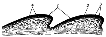
The surface layers of the epidermis of the skin of a lizard (like all other reptiles) become keratinized: the cells gradually die, filling with a horny substance - keratohyalin. Thickening of the stratum corneum occurs in small areas - scales, between which the stratum corneum is very thin, so the flexibility of the skin (and the whole body) is maintained. The shape of scales on different parts of the body of the same animal can vary significantly. U different types the shape, arrangement and number of scales are usually more or less different, so these features are widely used in the taxonomy of reptiles.
:
1 - stratum corneum (scales), 2 - epidermis, 3 - corium, 4 - pigment cells
The agama's head is covered with small, irregularly shaped scales; some other lizards have rather large horny scutes on their heads, arranged in a strictly defined order. On the upper surface of the head paired external nostrils are visible, opening into oral cavity the so-called internal nostrils, or choanae. The eyes are covered with moving eyelids; There is a nictitating membrane in the back corner of the eye. Behind the eyes are the ear openings, covered at some depth by the eardrum.
The elongated body of the agama is also covered with horny scales - small, irregularly shaped scales on the dorsal side and rows of larger scutes on the belly. At the posterior end of the body, at the border with the caudal region, between the abdominal scutes, there is a slit-like opening of the cloaca.
The tail scales of the Caucasian agama form double rings; in other lizards the arrangement of the tail scales is different.
The five-fingered limbs of lizards, like other reptiles, end in horny formations - claws.
The skin of lizards, like all reptiles, is dry, which is due to the absence of mucous glands. Skin glands are present in small numbers and are located only in a few areas specific to this species. They secrete a thick fat-like secretion and have special functions, most likely related to leaving an odorous trace that facilitates the formation of pairs during reproduction. In the agama, a group of such glands is clearly visible in the posterior part of the abdomen; their secretion in the form of a “waxy” coating covers the scales in this area. This accumulation of glands is especially well expressed in males.
General topography of internal organs
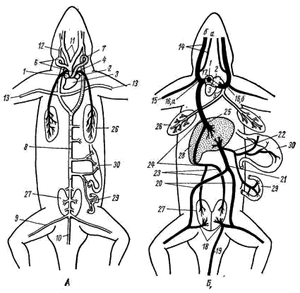
Circulatory system. The heart is located on the ventral side of the front of the chest cavity. Like amphibians, the heart of lizards is three-chambered: it consists of two atria - right and left - and one ventricle.
.
A - arterial system; B - venous system
(vessels with arterial blood are shown in white,
dotted line - with mixed and black - with venous blood):
1 - right atrium, 2 - left atrium, 3 - ventricle, 4 - pulmonary artery,
5 - pulmonary vein, 6 - right aortic arch, 7 - left aortic arch, 8 - dorsal aorta,
9 - iliac artery, 10 - caudal artery, 11 - carotid artery, 12 - carotid duct,
13 - subclavian artery, 14 - jugular veins(a - internal, b - external),
15 - subclavian vein, 16 - anterior vena cava (a - right, b - left),
17 - venous sinus, 18 - renal portal vein, 19 - tail vein, 20 - pelvic vein,
21 - abdominal vein, 22 - portal vein of the liver, 23 - renal vein, 24 - posterior vena cava,
25 - hepatic vein, 26 - lung, 27 - kidney, 28 - liver, 29 - intestines, 30 - stomach
The ventricle of the heart is divided by an incomplete, so-called horizontal septum, into two cavities: a smaller ventral (more precisely, ventrolateral), located down and to the right of the septum, and a large dorsal (dorsolateral) - up and to the left of the septum. The left atrium opens into left side the dorsal cavity of the stomach, and the right atrium - into the right part of the same cavity, in the area of the free edge of the septum. The dorsal cavity is divided into separate chambers by numerous muscle ridges. One of them, the most developed, is the so-called vertical septum, which divides the dorsal cavity of the ventricle into two halves - left and right. Due to this structure, complete mixing of arterial and venous blood does not occur in the ventricle of the reptile heart. When the atria contract, arterial blood expelled from the left atrium collects mainly in the left part of the dorsal cavity of the ventricle; venous blood from the right atrium enters the right half of the dorsal part of the ventricle and, flowing around the edge horizontal partition, collects in the ventral part of the ventricle. Only in the right half of the dorsal part of the ventricle does arterial and venous blood mix.
The arterial cone characteristic of amphibians in reptiles is reduced, and the main arterial trunks of the systemic and pulmonary circulation depart from the ventricle independently. Moreover, unlike amphibians, in which three pairs of arterial trunks depart from the arterial cone, in reptiles only three unpaired vessels begin in the heart: the pulmonary artery and two (right and left) aortic arches.
The pulmonary artery arises from the ventral (venous) part of the ventricle and soon divides into two branches carrying blood to the right and left lungs. Venous blood moves through the pulmonary arteries. Oxygenated arterial blood returns to the heart through the pulmonary veins. The right and left pulmonary veins merge into one unpaired vessel that flows into the left atrium. The entire system of the vessels considered makes up the pulmonary (pulmonary) circulation.
The vessels of the systemic circulation also begin in the ventricle of the heart. The right aortic arch departs from its left dorsal (arterial) part, and to the right of it, in the area of the free edge of the horizontal septum, the left aortic arch. According to the location of the origin of these vessels in the ventricle, predominantly arterial blood enters the right aortic arch, while mixed blood (arterial with an admixture of venous) enters the left one. Both arches of the aorta go around the heart and on the dorsal side behind it they unite into the unpaired dorsal aorta, which sends numerous vessels to various organs of the body. In the region of the hind limbs, the dorsal aorta branches into two large iliac arteries, which carry blood to the limbs, and the caudal artery.
The carotid arteries depart from the right aortic arch with a short, immediately bifurcating common trunk. Both carotid arteries, initially running parallel to the ascending branches of the aortic arches, carry blood to the head above the place where the aortic arches turn upward (downward from the observer) and each carotid artery sends back the carotid duct, which flows into the right or left aortic arch, respectively.
All of the listed vessels are quite clearly visible on a freshly killed lizard. If you carefully prepare the right aortic arch, then approximately in the middle between the place of its rotation and the place of confluence of the aortic arches, at the level of the posterior end of the heart, you can see the subclavian arteries extending from it and going to the forelimbs. Thus, in reptiles, unlike amphibians, the carotid and subclavian arteries arise asymmetrically - only from the right aortic arch. Thanks to this, the most oxygen-rich blood enters the head and forelimbs.
Venous blood from the head it collects into large paired jugular veins, which, merging with the less noticeable subclavian veins coming from the forelimbs, form the paired anterior vena cava. The anterior vena cava drains into the sinus venosus, which communicates with the right atrium. In lizards, the venous sinus, like in most reptiles, is weakly expressed.
From the back of the body, venous blood enters the heart in two ways. The veins carrying blood from the hind limbs form short paired portal veins of the kidneys, with each of which merge the branches of the divided unpaired caudal vein. These vessels can usually be seen only on injected preparations. Through the portal veins of the kidneys, blood enters the capillary system - the portal system of the kidneys.
Most of the blood from the posterior part of the body flows through fairly large paired pelvic veins, which, merging, form the unpaired abdominal vein, carrying venous blood to the liver. Venous blood from the intestine flows through several veins, merging into the azygos portal vein of the liver. In the liver or before entering it, the hepatic portal vein merges with the abdominal vein, and this common vessel immediately splits into a system of hepatic capillaries. Consequently, as in amphibians, the portal system of the liver is formed by two veins: the abdominal and portal liver.
From the portal system of the kidneys, blood collects in the paired renal veins, which merge into the large unpaired posterior vena cava. The posterior vena cava penetrates the liver (without sending vessels into it) and flows into the venous sinus. From the portal system of the liver, blood is collected through the capillary system into the short hepatic vein, which flows into the posterior vena cava at the anterior edge of the liver.
Respiratory system. The lizard's respiratory tract begins with the external nasal openings - the nostrils. Next, the air through the nasal passage and internal nostrils - choanae - enters the oral cavity. In the depths of the oral cavity, slightly in front of the esophagus, is the larynx, consisting of three cartilages. It is equipped with special muscles and is connected to the sublingual apparatus. From the oral cavity, the inhaled air through the larynx enters the trachea, a rather long tube in the walls of which there are ring-shaped cartilages that prevent it from collapsing. The trachea runs along the neck and in the chest cavity, approximately at the level of the heart, and divides into two short bronchi that enter the lungs.
The lungs are thin-walled hollow sacs. Compared to the lungs of amphibians, lizards have a more complex internal structure: their internal walls, in which the capillaries branch, have a spongy structure, which significantly increases the overall respiratory surface of the lungs.
The lungs are the only respiratory organ of reptiles. The skin of these animals is dry, covered with horny scales and keratinized epithelium and does not participate in respiration. The act of breathing in lizards occurs through expansion and contraction chest under the influence of special muscles.
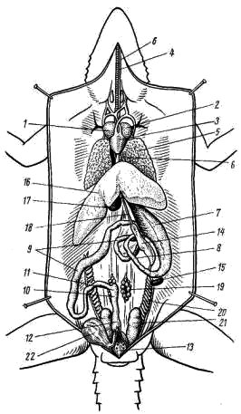
:
1 - right atrium, 2 - left atrium, 3 - ventricle, 4 - trachea, 5 - lung,
6 - esophagus, 7 - stomach, 8 - duodenum, 9 - small intestine,
10 - large intestine, 11 - rudimentary blind outgrowth of the intestine, 12 - rectum,
13 - cloacal cavity, 14 - pancreas, 15 - spleen, 16 - liver,
17 - gallbladder, 18 - bile duct, 19 - ovary, 20 - oviduct,
21 - kidney, 22 - bladder
Digestive system. In the oral cavity there is a flat tongue that tapers anteriorly; it helps in capturing and swallowing prey. Many lizards and snakes have a thin and long tongue, forking at the end. It is very mobile, can protrude quite far from the mouth and also serves as an organ of touch: lizards and snakes use it to feel objects in front of them. In addition, when the tongue is retracted into the mouth, its tips fall into special recesses equipped with sensory nerve endings - the Jacobson organ, which perceives chemical irritations from particles adhering to the tongue.
At the posterior end of the oral cavity behind the laryngeal fissure is the opening of the esophagus. The esophagus, in the form of a muscular, extensible tube, stretches along the neck above the trachea and in the front abdominal cavity flows into the stomach. From the posterior end of the stomach, the duodenum runs forward parallel to it, passing into small intestine. The boundary between the duodenum and small intestine is the first intestinal flexure (the place where the intestine turns back). The small intestine makes several bends and continues into the large intestine. At the border of the small and large intestines there is a small blind outgrowth - the rudiment of the cecum. The posterior part of the colon is the rectum. In lizards, the colon and rectum are separated by a poorly visible narrowing. The rectum opens into the cloaca and through the cloacal fissure to the outside.
Between the stomach and duodenum there is an elongated compact pancreas. Next to the stomach, towards the end of it, there is a small elongated reddish (on fresh material) spleen. The entire anterior part of the abdominal cavity is occupied by a large liver with several lobes. on her inside the gallbladder is located. The bile duct extending from it runs along the pancreas and flows into the beginning duodenum. The bile duct becomes more visible if you lightly press the gallbladder with tweezers and thus push some of the bile into the duct.
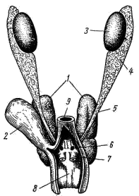
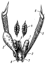
Genitourinary system. Unlike previously studied classes, reptiles in adulthood have not trunk (mesonephric) kidneys, but pelvic (metanephric) kidneys. They are located in the very back of the abdominal cavity and are covered by the pelvic bones. Along each kidney runs a ureter, which opens into the cloaca. The ureters of lizards, like other reptiles, are formed simultaneously with the development of the metanephric kidney as thin-walled protrusions of the posterior part of the Wolffian canals. The bladder extends from the abdominal wall of the cloaca in the form of a thin-walled blind outgrowth.
:
1 - kidney, 2 - bladder. 3-testis, 4 - epididymis, 5 - vas deferens,
6 - urogenital opening, 7 - copulatory sac, 8 - cloaca cavity,
9 - rectum
The male gonads - paired testes - are suspended on the mesentery in the posterior dorsal part of the abdominal cavity. The testes, with the help of the seminiferous tubules, are closely connected with the appendages of the testes, from which the vas deferens extend. Just before entering the cloaca, the vas deferens merge with the ureters and open into the cloaca with common openings. The appendages of the testis are the remains of the anterior section of the trunk (mesonephric) kidney, and the vas deferens are homologous to the excretory duct of this kidney - the Wolffian canal. Müllerian canals do not develop in males. In the side walls of the cloaca in males there are two hollow outgrowths that can turn out through the cloaca opening. They play the role of copulatory organs.
:
1 - kidney, 2 - bladder, 3 - urinary opening, 4 - ovary, 5 - oviduct,
6 - funnel of the oviduct, 7 - genital opening, 8 - cloaca cavity, 9 - rectum
The female gonads are paired ovaries, suspended in the abdominal cavity on the mesentery and do not have a direct connection with the excretory ducts. Mature eggs fall into the body cavity and are then captured by the oviduct funnel, which opens at the front of the body cavity. The oviducts, homologous to the Müllerian canals, open into the cloaca with independent (separate from the ureters) openings. The lower sections of the oviducts in lizards are often expanded and are then called the “uterus”. Wolffian canals are reduced in females.
Digestive system reptiles are more complex in structure compared to amphibians. The peculiarities of its structure in different species are determined type of food And way of obtaining food.
Majority reptiles carnivorous– their diet includes small terrestrial and aquatic animals, especially insects. However, larger vertebrate species are also eaten - fish, small amphibians, mouse-like rodents, and birds. Aquatic turtles, mainly feed on invertebrates and fish, but also consume algae. Reptiles-predators(crocodiles, boa constrictors, monitor lizards) attack larger prey; Often their victims are mammals - ungulates, waterfowl, monkeys and other animals.
In the food supply of reptiles, there is also narrow specialization: A number of species of tree snakes (egg snakes) feed exclusively eggs birds, and sea snakes - cephalopods. Herbivorous reptiles form a much smaller group. These species include: tropical agamas and marine iguanas that feed algae. Turtles, both terrestrial and many aquatic, also prefer vegetable food, but their “vegetarianism” is not absolute - they can also include small animals in their diet.
Oral cavity reptiles are well separated from throats and is partially separated from nasopharynx emerging secondary bony palate(in crocodiles and turtles it is fully formed). At the bottom of the oral cavity there is a well-developed language, which is attached by its base to its rear part. The tongue is equipped own muscles and has different shapes. With the help of a long tongue, which has easily extensible muscles and an adhesive substance, chameleons catch insects.
In lizards and snakes, the tongue is forked at the end and connected to Jacobson's organ, located on the palate and performing analyzer function in search of food. It is paired and looks like two holes, at the bottom of which there is sensitive epithelium And nerve endings.
In the oral cavity there are salivary glands, the secretion of which is used to moisten food. Some species of lizards and snakes have specialized glands producing toxic substances that immobilize prey.
The organ of prey capture in many reptiles is jaws, supplied with spicy conical teeth. The bases of the teeth grow into the jaw and pterygoid bones. In snakes, the ends of the teeth are bent slightly back, which ensures reliable retention of prey. A significant number of snakes have poisonous teeth, connected by channels with poisonous glands. The teeth of crocodiles sit in special formations of the jaws - alveoli. Most reptiles swallow their prey whole, but crocodiles and turtles swallow their food in chunks. Turtles, having no teeth, bite off food with the sharp edges of their jaws.
The oral cavity is followed by short throat, which goes into narrow esophagus(Fig. 46) . In many species that swallow large prey (snakes), it has strong and extensible muscles.
Stomach well demarcated, equipped with thick muscular walls and attached by a fold of peritoneum to the dorsal side of the body cavity. Intestines relatively long, clearly differentiated into sections - small, thick and rectum. To the anterior section small intestine – duodenum The ducts of the pancreas and large liver, which has a gallbladder, flow into it. On the border between the small and large intestines in many reptiles there is rudimentary cecum(it is well developed in herbivorous land turtles). Straightintestine has access to cloaca.
Respiratory system.
Reptiles breathe light, which are located in the thoracic region in the form of paired formations. Dermal breathing practically absent due to the dense horny covering of the body. Lungs most species are preserved cellular structure(Fig. 47), however, the number of cells and the total area of gas exchange are much larger than in amphibians. The structure of the lungs, their shape and total volume differ in reptiles of different systematic groups.
Right lung in snakes much larger than the left one amphisbaenus- vice versa. U lizards and snakes the inner surface of the lungs has folded cellular structure, which helps to increase the gas exchange surface. This structure is ensured by special outgrowths ( crossbars), extending from the internal walls of the organ. Connecting with each other, they form camera system increasing the respiratory surface.
Lungs of higher reptiles (crocodiles, turtles) have spongy structure, close in structure to the lung tissue of birds and animals. Special finger-like projections located in posterior region lungs in chameleons (Fig. 47 E), a number of lizards and snakes. They do not participate in gas exchange, but play the role of an additional air reservoir when absorbing food, diving into water and determine the hissing of reptiles.
Breathing mechanism in most reptiles chest, by expansion and contraction of the chest, which is achieved by contraction intercostal and abdominal muscles. When the chest expands, air is drawn in through external nostrils, choanae, oral cavity and laryngeal fissure into the laryngeal chamber. Next he gets into trachea, the initial part of which is supported by unpaired cricoid and arytenoid cartilages. From the back of the tracheal tube extend two bronchi, which branch in the lungs into smaller formations. The walls of the bronchi also have ring-shaped attachments.
In reptiles that do not have a chest (snakes, legless lizards), the flow of air into the respiratory system ensures, like amphibians, oropharyngeal mechanism. Turtles, whose chest is immovably connected by a shell, breathe thanks to the work whole muscle system – shoulder, abdominal, pelvic and lateral longitudinal bodies that increase the volume of the peripulmonary space and ensure that air is drawn inward.
The movement of the neck and forelimbs also contributes to the pumping of air into the lungs. In aquatic turtles, gas exchange takes place outgrowths of the pharynx and cloaca(“anal blisters”), where a network of capillaries branches abundantly. Fetal breathing occurs due to capillaries of the yolk sac and one of the embryonic membranes - allantois.
circulatory system, Compared to amphibians, it has undergone significant changes, which take place both in the structure of the heart and the vascular system. Heart three-chambere – two atria, separated full partition, And one ventricle. Venous the sinus is fused with the right atrium; there is no conus arteriosus. Both atria open into the ventricle independent holes.
The reverse flow of blood through them is prevented valves from semilunar folds. In the ventricle there is incomplete septum, which, when the heart contracts, approaches the atria. This contributes to a more complete division of blood into arterial and venous. U crocodiles partition full, however, with an opening, so the blood mixes, although not to the same extent as in many other reptiles.
Originates from different parts of the ventricle three arterial vessels with different blood content (Fig. 48 A). From left its parts originate right aortic arch, separating sleepy And subclavian arteries, carrying oxygenated blood to the head and forelimbs. From right side the ventricle departs pulmonary artery, which is divided into right and left arteries, carrying venous blood for oxidation to the lungs.
From middle part the ventricle originates left aortic arch with mixed blood. After going around the heart, the left and right aortic arches merge to form dorsal aorta, from which smaller vessels depart for blood supply to internal organs, trunk muscles and hind limbs.
Venous system reptiles are very similar to those of amphibians.
Venous blood from the head collects in paired jugular veins, which, merging with subclavian vessels, form anterior vena cava, flowing into the right atrium (Fig. 48 B). From the back of the body, venous blood flows through the azygos tail vein, giving rise to paired pelvic veins When combined with ischial veins are formed iliac veins.
The latter, separating the short ones renal portal veins, merge into unpaired abdominal vein, which, having received blood from the vessels coming from the intestines, forms in liver portal system. The capillaries of the portal system of the liver unite into hepatic veins, flowing into posterior vena cava. This large vessel is formed by the fusion portal veins of the kidneys, passes, without branching, through the liver and carries blood to the right atrium.
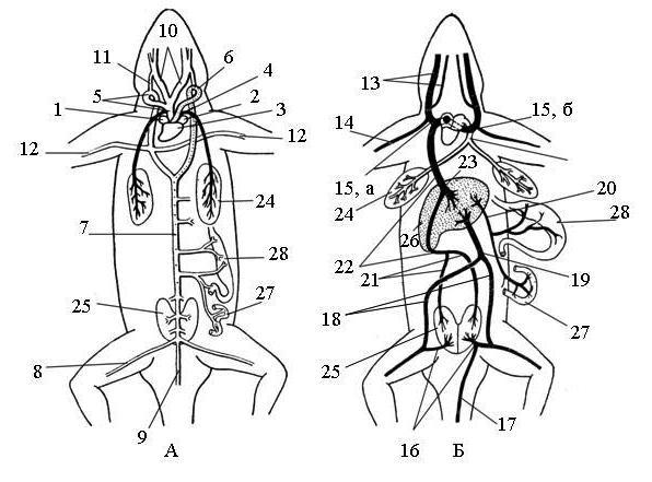
Fig.48. Scheme circulatory system Caucasian agama
(A – arterial, B – venous):
1 – right atrium, 2 – left atrium, 3 – ventricle, 4 – pulmonary artery, 5 – right aortic arch, 6 – left aortic arch, 7 – dorsal aorta, 8 – iliac artery, 9 – caudal artery, 10 – carotid artery , 11 – carotid duct, 12 – subclavian artery, 13 – jugular veins, 14 – subclavian vein, 15 – anterior vena cava (a – right, b – left), 16 – renal portal vein, 17 – tail vein, 18 – iliac vein vein, 19 - abdominal vein, 20 - hepatic portal vein, 21 - renal vein, 22 - posterior vena cava, 23 - hepatic vein, 24 - lung, 25 - kidneys, 26 - liver, 27 - intestines, 28 - stomach
Excretory system (Fig. 49) presented pelvic kidneys (metanephros), which are characteristic of all higher vertebrates (amniotes) and to the greatest extent provide the function of excretion in the terrestrial environment. They develop from special formations that form behind trunk kidneys, functioning at the embryonic stage and reducing towards the end of embryonic development. Simultaneously from the posterior part of the mesonephric ducts (Wolf channels) are being formed ureters.
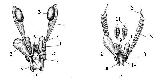
Rice. 49. Genitourinary system of the male (A) and female (B) Caucasian agama:
1 – kidney, 2 – bladder, 3 – testis, 4 – epididymis, 5 – vas deferens, 6 – male urogenital opening, 7 – copulatory sac, 8 – cloacal cavity, 9 – rectum, 10 – female urinary opening, 11 - ovary, 12 - oviduct, 13 - funnel of the oviduct, 14 - genital opening of the female.
Metanephric kidneys in the form of compact bodies are located in the pelvic region and differ from the trunk kidneys not only in location, but also microstructure. In the kidney complicated structure nephron tubules that contribute allocation exchange products and reverse suction water and salts.
Each tubule consists of four departments endowed with special functions. In its initial sections (proximal, intermediate and distal ) metabolic products are released and water and substances dissolved in it that are beneficial to the body are reabsorbed. These include sugars, vitamins, amino acids etc.
Last (collective) department withdraws final products of exchange in central renal canal, passing outside the kidney into ureter. As a result of an active process reabsorption, occurring in the kidney tubules, up to 90–95% of the substances from the original filtrate are returned to the bloodstream. This feature of the kidneys is especially important when reptiles live in arid conditions, where moisture deficiency has a dramatic effect and saving water in the body is crucial for survival.
Concentrated urine flows down the ureters into cloaca And bladder, where water reabsorption processes also take place, resulting in
Urine is released out in a very concentrated form. The main excretion product is uric acid, which does not require large amounts of water to remove. In many reptiles, urine looks like a suspension of small crystals (“white urine”).
In a number of reptiles living in sea water(sea turtles, iguanas), yes additional education, helping to remove excess salts from the body. They are located in the orbit of the eye (turtles) or near the nasal cavity (marine iguanas) and represent "salt glands" the secretion of which contains up to 7% salts - twice as much as in the environment.
Reproduction system.
Reptiles dioecious; the exception is one type of snake - island bothrops, which is hermaphrodite. Reptiles reproduce in different ways. Crocodiles, monitor lizards, pythons lay eggs. Common viper, spindle viper, viviparous lizard, sea snakes, sand boas are characterized by ovoviviparity.
Present live birth inherent in some chameleons, sea snakes, skinks and a number of other species. Fertilization only internal, with availability copulatory organs(only tuaterias don’t have them) . Snakes and lizards have paired copulatory organs; unpaired organ - in crocodiles and turtles.
Sex glands paired and located near the spine (Fig. 49). Testes represent oval bodies with smooth structure. Adjacent to them appendages, formed at trunk reduction kidneys Pass through the appendages seminiferous tubules, coming from the testes to genital ducts. Their function is performed Wolff channels, which near the cloaca flow into the lower part of the ureters.
Ovaries have grainy structure. Each oviduct (müllerian canal) opens funnel, supplied ciliated epithelium, into the body cavity near the ovary. The opposite end of the oviduct has an outlet into cloaca. Fertilization of an egg that enters the ovary occurs in its upper section.
As it passes through the oviduct, the zygote is covered successively protein, fibrous and shell shells. Around the developing embryo, special embryonic membranes(amniotic, serous and allantois), which additionally protect it from mechanical damage, ensure moisture retention, respiration and excretion.
Central nervous system and sensory organs.
Like other vertebrates , central nervous system reptiles consist of head and spinal departments
Brain has five departments, is covered with a dense pigmented shell on top and, in comparison with proto-terrestrial animals, has a number of progressive signs.
Anterior section brain large size due to growth striatum and medullary vault, in which, along with archipallium, there is a rudiment of the secondary arch - neopallium. The hemispheres of the brain are closely adjacent to each other, but their surface does not have convolutions. Olfactory department well expressed; through a long olfactory tract hemispheres connect with olfactory shares(Fig. 50).
Diencephalon covered from above by the forebrain and midbrain. IN roof areas contains two formations – pineal gland(endocrine gland) and parietal organ, performing a photosensitive function. The anterior part of the parietal organ is similar in structure to the lens of the eye, and its posterior part is pigmented and equipped with sensory cells. At the bottom diencephalon present funnel, above which are optic nerves and their cross (chiasma). Attached to the bottom of the funnel pituitary, part of the body's general endocrine system.
Midbrain well developed. It consists of two large front hills - optic lobes and small posterior colliculi. On the side there is bend brain, characteristic of all amniotes. The midbrain contains the main center processing visual information. Together with the anterior and intermediate parts of the brain, it determines behavior system reptiles.
Cerebellum presented in the form of a semicircular fold covering the anterior part of the medulla oblongata. Its relatively large dimensions reflect the significant activity of reptiles and the complexity of their movements.
Medulla oblongata has the appearance of a cord, wide in front and tapering at the back. At the site of its transition to the spinal cord there is second bend, characteristic of all higher vertebrates. The medulla oblongata is coordination center motor activity and a number of other vegetative functions - breathing, blood supply, digestion, etc. In turn, his activities are closely linked to the work forebrain hemispheres. Departs from the brain 11 pairs of nerves.
Spinal cord characterized by a clear division white matter(conducting pathways) and gray matter(nerve cells), which indicates increased coordination of its activity on the part of the brain. In the area of the limb girdles, nerve plexuses, enhancing the innervation of the muscles of the limbs.
Sense organs reflect a complex and diverse way of life and provide essential information about the environment.
Organvision plays a significant role in the lives of most reptiles. He is represented by paired eyes with moving eyelids And nictitating membrane, which protect the eyes from mechanical damage and drying out. This is facilitated by lacrimal glands. In snakes, geckos, skinks and a number of other reptiles, the eyelids grow together, creating a single cover and an unblinking gaze. Species that are predominantly nocturnal have vertical pupil. In some species (chameleons, some agamas and iguanas), the eyes can move independently of each other, which increases the angle of vision and allows for better orientation when catching prey.
Accommodation of the eye determined by the movement of the lens and changes in its shape using ciliary muscle. There is a special growth in the area of the back wall of the eyeball ("crest"), providing nutrition to the eye through an abundant network of blood capillaries. Black and white vision characteristic of nocturnal species, and colored – diurnal animals. Vision is determined by the presence of a certain amount rods and cones. Inhabitants of places with bright lighting have peculiar light filters, consisting of fat droplets, often pigmented.
Organhearing , like amphibians, consists of two sections - middle and inner. Average the ear includes one auditory ossicle - stirrup and covered eardrum. In internal ear separated from the middle ear round window, there is a well-developed, bag-shaped snail. This formation is an apparatus for recognizing incoming sound signals.
Organssense of smell paired The basis of each of them is nasopharyngeal passage, coming from the external nostrils and connecting to the oral cavity choanae. The middle part of the nasopharyngeal passage is divided into two sections - upper (olfactory) and lower (respiratory).
The olfactory department has from the inside folded structure, which helps to increase the surface area that perceives odors. On the palatal part of the oral cavity of reptiles there is two recesses (Jacobson organ), where microparticles of the object being examined and possible food are introduced for analysis with a forked tongue. In general, the sense of smell of reptiles is much more advanced than that of amphibians, and, together with vision and hearing, performs an important informational role.
Tactile function determined by the presence sensory hairs – derivatives keratinized skin cells, which are located along the edges of the horny scales.
A number of species (pit snakes, African vipers, pythons) have a special sensory organ - thermolocation , providing search for prey by temperature difference own body and warm-blooded animals, which they mainly hunt. This paired organ is located in the front of the head - on the upper labial scutes (pythons), in the recesses between the nostrils and eyes (pit snakes, African vipers). Each hole is covered on the outside with a transparent film and separated from the internal cavity thin membrane. Information from outside enters the cavity through a thin channel and is captured by those lying on the bottom thermoreceptors. The organ is innervated by the branch trigeminal nerve.