Structurally, the anatomical unit of bone. Bone as an organ (bone structure)
Most adult human bones consist of lamellar bone tissue. It forms a compact substance located along the periphery, and a spongy substance - the masses of bone crossbars in the middle of the bone.
Compact substance substantia compacta, bones form diaphyses tubular bones, in the form of a thin plate, covers the outside of their epiphyses, as well as spongy and flat bones built from spongy substance. The compact substance of the bones is penetrated by thin channels in which blood vessels and nerve fibers. Some channels are located predominantly parallel to the surface of the bone ( central, or Haversian, canals), others open on the surface of the bone with nutrient openings (foramina nutricia), through which arteries and nerves penetrate into the thickness of the bone, and veins emerge.
The walls of the central (Haversian) canals are formed by concentric plates located around the central canal. Around one canal there are from 4 to 20 such bone plates, as if inserted into each other. The central channel, together with the plates surrounding it, is called osteon(Haversian system). Osteon is a structural and functional unit of compact bone substance (Fig. 2.2).
Spongy substance, substantia spongiosa, represented by interconnected trabeculae, forming a spatial lattice resembling a honeycomb. Its crossbars are not arranged randomly, but naturally, in accordance with functional conditions. The structural and functional unit of the spongy substance is the trabecular packet, which is a set of parallel bone plates within one trabecula and delimited from each other by a cleavage line. Bone cells contain bone marrow– organ of hematopoiesis and biological defense of the body. It is also involved in nutrition, development and bone growth. In tubular bones, the bone marrow is also located in the canal of these bones, called, therefore, bone marrow cavity, cavitas medullaris . Thus, all the internal spaces of the bone are filled with bone marrow, which forms an integral part of the bone as an organ. There are red bone marrow and yellow bone marrow.
red bone marrow, medulla ossium rubra, has the appearance of a tender red mass consisting of reticular tissue, in the loops of which there are cellular elements that are directly related to hematopoiesis (stem cells), to immune system and bone formation (bone builders - osteoblasts and bone destroyers - osteoclasts), blood vessels and blood elements give the bone marrow its red color.
Yellow bone marrow medulla ossium flava, owes its color to the fat cells from which it is composed.
The distribution of compact and cancellous substance depends on bone function. The compact substance is found in those bones and in those parts of them that primarily perform the function of support (stand) and movement (levers), for example, in the diaphysis of tubular bones. In places where, with a large volume, it is necessary to maintain lightness and at the same time strength, a spongy substance is formed, for example, in the epiphyses of tubular bones (Fig. 2.2).
Figure 2.2. Femur.
a – structure of the femur on a cut; b – the crossbars of the spongy substance are located not randomly, but regularly; 1 – pineal gland; 2 – metaphysis; 3 – apophysis; 4 – spongy substance; 5 – diaphysis; 6 – compact substance; 7 – bone marrow cavity.
The entire bone, except for the points of connection with the bones (articular cartilage), is covered with a connective tissue membrane - periosteum, periosteum (periosteum). This is a thin, strong connective tissue film of pale pink color that surrounds the bone from the outside, consisting in adults of two layers: the outer fibrous (fibrous) and the inner bone-forming (osteogenic, or cambial). It is rich in nerves and blood vessels, due to which it participates in the nutrition and growth of bone thickness.
Thus, the concept of bone as an organ includes bone tissue, which forms the main mass of the bone, as well as bone marrow, periosteum, articular cartilage and numerous nerves and vessels.
Chemical composition of bones complex. In a living organism, the bones of an adult human contain about 50% water, 28% organic and 22% inorganic substances. Inorganic substances are represented by compounds of calcium, phosphorus, magnesium and other elements. The organic substances of bone are collagen fibers, proteins (95%), fats and carbohydrates (5%). These substances give bones firmness and elasticity. With an increase in the proportion of inorganic compounds (in old age, in some diseases) the bone becomes brittle and brittle. The strength of bone is ensured by the physicochemical unity of inorganic and organic substances and the features of its design. The chemical composition of bones depends on age (organic substances predominate in children, inorganic substances in old people), the general condition of the body, functional loads, etc. With a number of diseases, the composition of bones changes.
The composition of fresh adult human bone includes water - 50%, fat - 16%, other organic substances - 12%, inorganic substances - 22%.
Defatted and dried bones contain approximately 2/3 inorganic and 1/3 organic matter. In addition, bones contain vitamins A, D and C.
Organic matter of bone tissue - ossein– gives them elasticity. It dissolves when boiled in water, forming bone glue. Inorganic bone matter is represented mainly by calcium salts, which with a small admixture of other mineral substances form hydroxyapatite crystals.
The combination of organic and inorganic substances determines the strength and lightness of bone tissue. So, with a low specific gravity of 1.87, i.e. not twice the specific gravity of water, the strength of bone exceeds the strength of granite. The femur, for example, when compressed along the longitudinal axis, can withstand loads of over 1500 kg. If a bone is fired, the organic substance burns out, but the inorganic substance remains and retains the shape of the bone and its hardness, but such a bone becomes very fragile and crumbles when pressed. On the contrary, after soaking in a solution of acids, as a result of which the mineral salts dissolve and the organic matter remains, the bone also retains its shape, but becomes so elastic that it can be tied into a knot. Consequently, the elasticity of the bone depends on ossein, and its hardness - on mineral substances.
The chemical composition of bones is associated with age, functional load, general condition body. The greater the load on the bone, the more inorganic substances there are. For example, the femur and lumbar vertebrae contain the largest amount of calcium carbonate. With increasing age, the amount of organic substances decreases, and inorganic substances increase. In young children there is comparatively more ossein; accordingly, the bones are highly flexible and therefore rarely break. On the contrary, in old age the ratio of organic and inorganic substances changes in favor of the latter. Bones become less elastic and more fragile, as a result of which bone fractures are most often observed in old people.
Classification of bones
Based on shape, function and development, bones are divided into three parts: tubular, spongy, mixed.
Tubular bones are part of the skeleton of the limbs, playing the role of levers in those parts of the body where large-scale movements predominate. Tubular bones are divided into long – humerus, bones of the forearm, femur, bones of the leg and short– bones of the metacarpus, metatarsus and phalanges of the fingers. Tubular bones are characterized by the presence of a middle part - diaphysis, containing a cavity (marrow cavity), and two expanded ends - epiphyses. One of the epiphyses is located closer to the body - proximal, the other is further from him – distal. The section of tubular bone located between the diaphysis and epiphysis is called metaphysis. The bone processes that serve to attach muscles are called apophyses.
Spongy bones are located in those parts of the skeleton where it is necessary to provide sufficient strength and support with a small range of movements. Among the spongy bones there are long(ribs, sternum), short(vertebrae, carpal bones, tarsus) and flat(skull bones, belt bones). Spongy bones include sesamoid bones (patella, pisiform bone, sesamoid bones of the fingers and toes). They are located near the joints, are not directly connected to the bones of the skeleton and develop in the thickness of the muscle tendons. The presence of these bones helps to increase the muscle's leverage and therefore increase its torque.
Mixed dice– this includes bones that merge from several parts that have different functions, structure and development (bones of the base of the skull).
Bone as an organ (bone structure).
Bone, os, ossis, as an organ of a living organism consists of several tissues, the most important of which is bone.
Chemical composition of bone and its physical properties.
Bone substance consists of two types chemicals: organic (Uz), mainly ossein, and inorganic (2/3), mainly calcium salts, especially phosphate of lime (more than half - 51.04%). If the bone is exposed to a solution of acids (hydrochloric, nitric, etc.), then the lime salts dissolve (decalcinatio), and the organic matter remains and retains the shape of the bone, being, however, soft and elastic. If the bone is fired, the organic substance burns out, and the inorganic substance remains, also retaining the shape of the bone and its hardness, but being very fragile. Consequently, the elasticity of bone depends on ossein, and its hardness on mineral salts. The combination of inorganic and organic substances in living bone gives it extraordinary strength and elasticity. They are convinced of this age-related changes bones. In young children, who have relatively more ossein, the bones are highly flexible and therefore rarely break. On the contrary, in old age, when the ratio of organic and inorganic substances changes in favor of the latter, bones become less elastic and more fragile, as a result of which bone fractures are most often observed in old people.
Bone structure.
The structural unit of bone, visible through a magnifying glass or at low magnification of a microscope, is osteon , i.e., a system of bone plates concentrically located around a central canal containing blood vessels and nerves.
Osteons do not adhere closely to each other, and the spaces between them are filled with interstitial bone plates. Osteons are not located randomly, but according to the functional load on the bone: in tubular bones parallel to the length of the bone, in spongy bones - perpendicular vertical axis, in flat bones of the skull - parallel to the surface of the bone and radially.
Together with the interstitial plates, osteons form the main middle layer of bone substance, covered from the inside (from the endosteum) by the inner layer of bone plates, and from the outside (from the periosteum) by the outer layer of the surrounding plates. The latter is penetrated by blood vessels coming from the periosteum into the bone substance in special perforating canals. The beginning of these canals is visible on the macerated bone in the form of numerous nutrient holes (foramina nutrfcia). The blood vessels passing through the canals ensure metabolism in the bone. Larger bone elements, visible to the naked eye on a cut or on an x-ray, are made up of osteons - crossbars of bone substance, or trabeculae. These trabeculae make up two types of bone substance: if the trabeculae lie tightly, then it turns out dense compact substance, substantia compacta. If the trabeculae lie loosely, forming bone cells between themselves like a sponge, then it turns out spongy, trabecular substance, substantia spongiosa, trabecularis (spongia, Greek - sponge).
The distribution of compact and cancellous substance depends on the functional conditions of the bone. The compact substance is found in those bones and in those parts of them that primarily perform the function of support (stand) and movement (levers), for example, in the diaphysis of tubular bones.
In places where, with a large volume, it is necessary to maintain lightness and at the same time strength, a spongy substance is formed, for example, in the epiphyses of tubular bones (Fig. 7).
The crossbars of the spongy substance are not arranged randomly, but regularly, also in accordance with the functional conditions in which a given bone or part of it is located. Since the bones experience a double action - pressure and muscle traction, the bone crossbars are located along the lines of compression and tension forces. According to the different directions of these forces, different bones or even parts of them have different structure. In the integumentary bones of the cranial vault, which primarily perform a protective function, the spongy substance has a special character that distinguishes it from other bones that carry all 3 skeletal functions. This spongy substance is called diploe, diploe (double), since it consists of irregularly shaped bone cells located between two bone plates - the outer, lamina externa, and the inner, lamina interna. The latter is also called vitreous, lamina vftrea, since it breaks when the skull is damaged more easily than the outer one.
Bone cells contain bone marrow - organ of hematopoiesis and biological defense of the body. It is also involved in nutrition, development and bone growth. In tubular bones, the bone marrow is also located in the canal of these bones, therefore called the medullary cavity, cavitas medullaris.
Thus, all the internal spaces of the bone are filled with bone marrow, which forms an integral part of the bone as an organ.
There are two types of bone marrow: red and yellow.
Red bone marrow, medulla ossium rubra (for structural details, see the histology course), has the appearance of a tender red mass consisting of reticular tissue, in the loops of which there are cellular elements that are directly related to hematopoiesis (stem cells) and bone formation (bone builders - osteoblasts and bone destroyers - osteoclasts). It is penetrated by nerves and blood vessels that, in addition to the bone marrow, supply the inner layers of the bone. Blood vessels and blood elements give bone marrow its red color.
Yellow bone marrow, medulla ossium flava, owes its color to the fat cells of which it is mainly composed.
During the period of development and growth of the body, when greater hematopoietic and bone-forming functions are required, red bone marrow predominates (in fetuses and newborns there is only red marrow). As the child grows, the red marrow is gradually replaced by yellow marrow, which in adults completely fills the medullary cavity of the tubular bones.
On the outside, the bone, with the exception of the articular surfaces, is covered with periosteum (periosteum).
Periosteum- this is a thin, strong connective tissue film of pale pink color, surrounding the bone from the outside and attached to it with the help of connective tissue bundles - perforating fibers that penetrate the bone through special tubules. It consists of two layers: the outer fibrous (fibrous) and the inner bone-forming (osteogenic, or cambial). It is rich in nerves and blood vessels, due to which it participates in the nutrition and growth of bone thickness. Nutrition is carried out by blood vessels penetrating in large numbers from the periosteum into the outer compact substance of the bone through numerous nutrient openings (foramina nutricia), and bone growth is carried out by osteoblasts located in the inner layer adjacent to the bone (cambium). The articular surfaces of the bone, free from periosteum, are covered by articular cartilage, cartilage articularis.
Thus, the concept of bone as an organ includes bone tissue, which forms the main mass of the bone, as well as bone marrow, periosteum, articular cartilage and numerous nerves and vessels.
Test questions for the lecture:
1. The concept of bone (hard) and connective tissue skeleton,
2. General overview human skeleton, classification of bones.
3. The structure of bone as an organ, periosteum, bone marrow.
4. Osteon structure: Haversian canals, bone plates; bone cells - osteoblasts, osteocytes, osteoclasts.
5. Bone structure; diaphysis, metaphysis, epiphysis, apophysis, compact and spongy substance.
6. Chemical composition of bone.
Lecture No. 5
Bone in x-ray image. The influence of labor and sports on the structure of the bones of a living person. The relationship between social and biological factors in the structure of bones.
Purpose of the lecture. Consider the structure of bone in the whole organism.
lecture plan:
1. Consider the x-ray anatomy of bones.
2. Consider the dependence of bone development on internal and external factors.
3. Reveal the structural and functional relationships between the active and passive parts of the musculoskeletal system.
4. Reveal the role of the Russian scientist P.F. Lesgaft in the study of the interdependence of the muscular and skeletal systems.
5. Consider the relationship between social and biological factors in the formation of the human skeleton.
X-RAY ANATOMY OF BONES.
On radiographs, compact and spongy substances are clearly distinguishable. The first gives an intense contrasting shadow, corresponding to the plane of the cortical layer, and in the area of substantia spongiosa the shadow has a network-like character (see Fig. 1).
Compact substance of the epiphyses of tubular bones and the compact substance of bones, built primarily from spongy substance (bones of the wrist, tarsus, vertebrae), has the appearance of a thin layer bordering the spongy substance. This thin cortical layer on the articular sockets appears thicker than on the articular heads.
In the diaphyses of tubular bones, compact the substance varies in thickness: in the middle part it is thicker, tapering towards the ends. In this case, between the two shadows of the cortical layer, a bone marrow cavity is noticeable in the form of some clearing against the background of the general shadow of the bone. If this cavity is not traced throughout its entire length, this indicates the presence of a pathological process.
X-ray contours of the compact substance of the diaphysis clear and smooth. At the attachment points of ligaments and muscles, the contours of the bone are uneven. Against the background of the cortical layer of the diaphysis, thin stripes of clearing are noticeable, corresponding to the vascular canals. They are usually located obliquely: in long tubular bones upper limb- closer and towards the elbow joint; in the long tubular bones of the lower limb - further and in the direction from knee joint; in short tubular bones of the hand and foot - closer and towards the end that does not have a true epiphysis.
Spongy substance on x-ray has the appearance of a looped network consisting of bone crossbars with enlightenments between them. The nature of this network depends on the location of the bone plates in a given area, according to the compression and tension lines.
Bone development. X-ray examination skeletal system becomes possible from the 2nd month of uterine life, when on the basis of cartilage or connective tissue ossification points appear.
Appearance ossification points easily identified on radiographs, and these points, separated by cartilage tissue, look like separate bone fragments. They can give rise to erroneous diagnoses of fracture, fracture or necrosis (death) of the bone. Because of this, knowledge of the location of bone nuclei, the timing and order of their appearance is extremely important in practical terms.
Therefore, we present ossification in all relevant places on the basis of data not from an anatomical study of corpses, but from x-ray anatomy (study of a living person).
In cases of non-fusion of accessory nuclei with the main part of the bone, they can remain for life in the form of independent, unstable or accessory bones. Their detection on an x-ray may lead to diagnostic errors.
All major ossification nuclei appear in the bones of the skeleton before the onset of puberty, called puberty. WITH the onset of puberty the fusion of the epiphyses with the metaphyses begins, i.e., the transformation of synchondrosis connecting the bone epiphysis with the bone metaphysis into synostosis. This is radiographically expressed in the gradual disappearance of clearing at the site of the metaepiphyseal zone, corresponding to the metaepiphyseal cartilage separating the epiphysis from the metaphysis. Upon the onset of complete synostosis, traces of the former synchondrosis cannot be determined (Fig. 1).
Aging of the skeletal system. In old age, the skeletal system undergoes significant changes. On the one hand, there is a decrease in the number of bone plates and bone loss (osteoporosis); on the other hand, excessive bone formation occurs in the form of bone growths (o s t e f i t o v) and calcification of articular cartilage, ligaments and tendons at the site of their attachment to the bone.
Accordingly, the X-ray picture of aging of the osteoarticular apparatus consists of the following changes, which should not be interpreted as symptoms of pathology (degeneration).
I. Changes caused by atrophy of bone substance:
1) osteoporosis (on the x-ray the bone becomes more transparent);
2) deformation of the articular heads (disappearance of their round shape, “grinding down” of the edges, appearance of “corners”).
II. Changes caused by excessive deposition of lime in the connective tissue and cartilaginous formations adjacent to the bone:
1) narrowing of the joint “X-ray” gap due to calcification of articular cartilage;
2) strengthening of the diaphysis relief due to calcification at the site of attachment of tendons and their fibrous sheaths;
3) bone growths - osteophytes , formed as a result of calcification of ligaments at the site of their attachment to the bone.
The described changes are especially clearly visible in the spine and hand. In the remaining parts of the skeleton, three main radiological symptoms of aging are observed: osteoporosis, increased bone relief and narrowing of joint spaces. For some people, these signs of aging are noticed early (30-40 years), for others - late (60-70 years) or not at all.
Summarizing the presentation of general data on the ontogenesis of the skeletal system, we can say that x-ray examination makes it possible to more accurately and deeply study the development of the skeleton in its functioning state than the study of only cadaveric material.
In this case, a number of normal morphological changes are noted:
1) the appearance of ossification points - main and additional;
2) the process of their synostosis with each other;
3) senile involution of bone.
The described changes are normal manifestations of age-related variability of the skeletal system. Consequently, the concept of “norm” cannot be limited only to an adult and considered as a single type. This concept must be extended to all other ages.
DEPENDENCE OF BONE DEVELOPMENT ON INTERNAL AND EXTERNAL FACTORS
The skeleton, like any organ system, is a part of the body that reflects various processes occurring in it. Therefore, many factors influence the development of the skeletal system.
Influence of internal factors. X-ray examination reveals a number of morphological changes in bones, depending on the activity of other organs. It is especially clearly determined by radiography connection between the skeletal system and the endocrine glands. The active activation of the gonads entails the onset of puberty, puberty . Before this, in the prepubertal period, the activity of other endocrine glands, the appendage of the brain - the pituitary gland, increases, the function of which is associated with the appearance of ossification nuclei. By the beginning of the prepubertal period, all the main ossification points appear, and there is a gender difference in the timing of their appearance: in girls 1-4 years earlier than in boys. The onset of the prepubertal period, associated with the function of the pituitary gland, coincides with the appearance of an ossification nucleus in the pisiform bone, which belongs to the category of sesamoid bones.
On the eve of puberty, other sesamoid bones also ossify, namely at the metacarpophalangeal joint of the first finger. The beginning of the pubertal period, when, in the words of the famous endocrine researcher Beadle, “the sex glands begin to play the main melody in the endocrine concert,” is manifested in the skeletal system by the onset of synostoses between the epiphyses and metaphyses, with the very first such synostosis observed in the first metacarpal bone. Therefore, based on its comparison with other data on sexual development (the appearance of terminal vegetation, the onset of menstruation, etc.), synostosis of the 1st metacarpal bone is considered an indicator of incipient puberty, i.e., an indicator of the onset of puberty; among St. Petersburg residents, synostosis of the first metacarpal bone occurs at the age of 15-19 years in boys and at 13-18 years in girls.
Full puberty, also receives a well-known reflection in the skeleton: at this time, synostoses of the epiphyses with metaphyses in all tubular bones end, which is observed in women aged 17-21 years, and in men - at 19-23 years. Since the end of the process of synostosis ends the growth of bones in length, it becomes clear why men who have puberty ends later than in women; in general, they are taller than women.
Considering this connection between the skeletal system and the endocrine system and comparing data on age characteristics skeleton with data on puberty and general development of the body, we can talk about the so-called “bone age”. Thanks to this, from the X-ray picture of some parts of the skeleton, especially the hand, it is possible to determine the age of a given individual or judge the correctness of his ossification process, which is of practical importance for diagnosis, forensic medicine, etc. Moreover, if the “passport” age indicates the number of years lived years (i.e. on the quantitative side), then the “bone” age to a certain extent indicates their qualitative side.
X-ray examination also reveals dependence of bone structure on condition nervous system , which, regulating all processes in the body, carries out, in particular, the trophic function of bone. At enhanced trophic function of the nervous system More bone tissue is deposited in the bone, and it becomes denser and more compact (osteosclerosis). On the contrary, when weakening of trophism bone loss is observed - osteoporosis. The nervous system also influences the bone through the muscles, the contraction of which it controls (as will be discussed below). Finally, the various parts of the central and peripheral nervous system determine the shape of the surrounding and adjacent bones. So, all vertebrae form a spinal canal around spinal cord. The bones of the skull form a bony box around the brain and take on the shape of the latter. In general, bone tissue develops around elements of the peripheral nervous system, resulting in the formation of bone canals, grooves and pits that serve for the passage of nerves and other nerve formations (nodes).
Bone development is also in very close depending on the circulatory system. The entire process of ossification from the moment the first bone nucleus appears until the end of synostosis takes place with the direct participation of vessels, which, penetrating into the cartilage, contribute to its destruction and replacement with bone tissue. In this case, bone plates (Haversian) are deposited in in a certain order around blood vessels, forming Haversian systems with a central channel for the corresponding vessel. Consequently, when bone arises, it is built around blood vessels. This also explains the formation of vascular canals and grooves in the bones at the places where arteries and veins pass and adjoin them.
Ossification and bone growth after birth also occurs in close dependence on blood supply. It is possible to outline a number of stages of age-related variability in bone associated with corresponding changes in the bloodstream (Fig. 2).
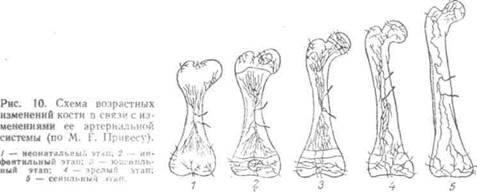
1. Neonatal stage , characteristic of the fetus (the last months of intrauterine development) and the newborn; the vascular bed of the bone is divided into a number of vascular regions (epiphysis, diaphysis, metaphysis, apophysis), which do not communicate with each other (closedness, isolation) and within which the vessels do not connect to each other, do not anastomose (terminal nature of the vessels, “limb”) .
2. Infantile stage , characteristic of children before the onset of synostosis; the vascular regions are still separated, but within each of them the vessels anastomose with each other and their terminal character disappears (“closedness” in the absence of a “limb”).
3. Juvenile stage , characteristic of young men, begins with the establishment of connections between the vessels of the epiphysis and metaphysis through the metaepiphyseal cartilage, due to which the closedness of the epiphyseal begins to disappear. metaphyseal and diaphyseal vessels.
4. Mature stage , characteristic of adults; synostoses occur, and all intraosseous vessels form unified system: They are not “closed” or “finite”.
5. Senile stage , characteristic of old people; the vessels become thinner and the entire vascular network becomes poorer.
On the shape and position of the bones affect to the inside, for which they form bone receptacles, beds, pits, etc.
The formation of the skeleton and organs refers to the beginning of embryonic life; during their development, they influence each other, which is why there is a correspondence between organs and their bone containers, for example chest and lungs, pelvis and its organs, skull and brain, etc.
The development of the entire skeleton must be considered in the light of these relationships.
Influence of external (social) factors on the structure and development of the skeleton. Unity of form and function in the structure of bones. Influencing nature in the process of labor activity, a person sets in motion his natural tools - arms, legs, fingers, etc. In the tools of labor, he acquires new artificial organs that complement and lengthen the natural organs of the body, changing their structure. And the man himself “...at the same time changes his own
 |
nature." Hence, labor processes have a significant impact on the human body as a whole, on its movement apparatus, including the skeletal system.
Reflects especially clearly on the skeleton muscle work. As shown experimental studies P.F. Lesgaft, the stronger the muscle work, the better the bone develops, and vice versa. At the sites of tendon attachment, protrusions (tubercles, processes,
roughness), and locally
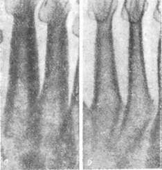
Rice. 3. Radiographs of the metatarsal bones.
places of attachment of the muscles of the ballerina (a) and sedentary workers (b).
attachments of muscle bundles - smooth or concave surfaces (pits).
RELATIONSHIP OF THE ACTIVE AND PASSIVE PART OF THE MUSCULOCAL SYSTEM
The more developed the muscles, the better the places of muscle attachment on the bones are expressed. This is why the bone relief, caused by the attachment of muscles, is more pronounced in an adult than in a child, and more pronounced in men than in women.
Prolonged and systematic muscle contractions, as occurs during exercise and professional work, gradually cause, through the reflex mechanisms of the nervous system, a change in metabolism in the bone, resulting in an increase in bone substance, called working hypertrophy (Fig. 3). This working hypertrophy causes changes in the size, shape and structure of bones, which are easily determined radiographically in living people.
Different professions require different physical work, which is associated with different degrees of participation of certain bones in this work.
Increased physical stress on the movement apparatus causes working hypertrophy of the bones, as a result of which their shape, width and length change, as well as the thickness of the compact substance and the size of the medullary space; The structure of the spongy substance also changes.
Width of bones. Thus, for loaders, the width of their bones, as their professional experience increases, reaches significantly larger sizes than for representatives of office work.
Research by P.F. Lesgaft identified a number of patterns in the relationship between the active and passive parts of the musculoskeletal system. They established:
1. Bones develop more strongly, the greater the activity of the surrounding muscles; with less load on the organs, they become thinner, longer, narrower and weaker.
2. The shape of the bones changes depending on the pressure of the surrounding organs (muscles, skin, eyes, teeth, etc.), they thicken and are directed towards the least resistance.
3. The shape of the bone also changes due to the pressure of the external parts; the bone grows more slowly due to increased external pressure, bending under the influence of one-sided action.
4. Fascia - thin membranes that cover and separate the muscles and are directly influenced by them, also exert lateral pressure on the bones.
5. Bones are active in relation to the shape of their structure (architecture), playing the role of racks or supports for surrounding organs.
RELATIONSHIP OF SOCIAL AND BIOLOGICAL IN BONE STRUCTURE
The bone is not a frozen model that does not change after its formation, as previously thought. This metaphysical view has been overcome modern anatomy, which considers the vital activity of bone, even in an adult, as a continuous exchange of substances with other tissues of the body, as a dialectical unity and struggle of two opposing processes - bone formation and bone destruction (resorption; resorptio - resorption). As a result of this struggle, there is permanent shift bone structures and its chemical composition; so, for example, the femur is completely renewed within 50 days. In this case, the bone is subject to a number of biological laws: adaptation (adaptation) to new living conditions, the unity of the organism and the environment, the unity of form and function, variability as a result of exercise or lack of exercise, the effect of mechanical compression of one part on another, etc. The morphological expression of these laws in relation to the skeleton is the restructuring of the bone structure (bone restructuring) in accordance with changing functional needs, about which already mentioned above.
This, in brief, is the “biological side” of the relationship between the social and the biological. As for the “social side”, the following must be kept in mind.
Various social factors (profession, lifestyle, diet, etc.) are associated with different physical activity, which determines the varying degrees of participation of certain bones in this work. The work of a professional worker requires a long stay of the body in one position or another (for example, a bent position over a machine or desk) or a constant change in body position in one direction or another (for example, bending the torso forward and throwing it back in carpenters). Therefore, the nature of the professional load and its volume determine the greater or lesser participation in the work of a given part of the skeleton and each bone separately and determine the different nature and degree of restructuring of its structure. When changing profession, bone restructuring is observed in the direction of increasing or weakening working hypertrophy, depending on the nature of the professional load. The growth of bones in length increases with favorable physical activity.
Bone aging occurs later in workers who have properly organized long-term physical labor, which does not cause premature wear of bone tissue.
The stated facts of individual variability of the skeletal system are due to both biological and social factors. Environmental irritants are perceived biologically by the body and lead to skeletal restructuring. The ability of bone tissue to adapt to changing functional needs through bone remodeling is the biological cause of bone variability, and the nature of the profession, the volume of professional load, the intensity of work, and lifestyle this person and other social factors are the social causes of this variability.
This is the relationship between the social and the biological in the structure of the skeleton. Knowing this relationship, it is possible to specifically influence the structure of the skeletal system by selecting appropriate physical exercise in work and sports and through changes in social conditions of life.
Test questions for the lecture:
1. X-ray anatomy of bones.
2. Dependence of bone development on internal and external factors.
3. Structural and functional relationships between the active and passive parts of the musculoskeletal system.
4. The role of the Russian scientist P.F. Lesgaft in the study of the interdependence of the muscular and skeletal systems.
5. The relationship between social and biological factors in the formation of the human skeleton.
Lecture No. 6
General arthrosyndesmology.
Purpose of the lecture. Consider the functional anatomical features various types bone connections.
lecture plan:
1. Consider the development of bone joints in phylogeny.
2. Consider the classification of bone connections.
3. Reveal the functional anatomy of syndesmoses.
4. Reveal the functional anatomy of synchrodrosis, synostosis, and semi-joints.
5. Consider the classification of joints according to the number of articular surfaces and the shape of the articular surfaces.
6. Consider the classification of joints according to the number of axes of motion.
7. Consider general characteristics combined joints and complex joints.
8. Consider the structure of the main and auxiliary elements of the joints.
9. Reveal the basic principles of joint biomechanics.
10. Reveal the functional and morphological features of the spinal column as a whole.
11. Reveal the functional and morphological features of the pelvis as a whole.
12. Reveal the functional and morphological features of the foot as a whole.
DEVELOPMENT OF BONE JOINTS IN PHYLOGENESIS
The initial form of bone connection is their fusion with the help of connective or (later) cartilaginous tissue. However, this continuous method of connecting the bones limits the range of movement. With the formation of bone levers of movement in the tissue intermediate between the bones due to the resorption of the latter, cracks and cavities appear, resulting in new look connection of bones - discontinuous, articulation. The bones began not only to connect, but also to articulate, joints were formed that allowed the bone levers to produce extensive movements. Thus, in the process of phylogenesis, 2 types of bone connections developed: the initial one was continuous, continuous with a limited range of movements, and the later one was discontinuous, allowing extensive movements. Reflecting this phylogenetic process in human embryogenesis, the development of bone joints goes through these 2 stages. Initially, the skeletal rudiments are continuously interconnected by layers of mesenchyme. The latter turns into connective tissue, from which the apparatus that connects the bones is formed. If the areas of connective tissue located between the bones turn out to be solid, then a continuous continuous connection of bones will result - fusion, or synarthrosis. If a cavity is formed inside them by resorption of connective tissue, then another type of connection occurs - cavitary, or discontinuous - diarthrosis.
Thus, according to development, structure and function, all bone joints can be divided into 2 large groups:
1. Continuous connections- synarthrosis(BNA) - earlier in development, immobile or sedentary in function.
2. Discontinuous connections - diarthrosis(BNA) - later in development and more mobile in function.
There is a transition between these forms - from continuous to discontinuous or vice versa. It is characterized by the presence of a small gap that does not have the structure of a real articular cavity, as a result of which this form is called semi-joint - symphysis, symphysis (BNA).
Bone, os, ossis, as an organ of a living organism consists of several tissues, the most important of which is bone.
Chemical composition of bone and its physical properties.
Bone substance consists of two types of chemical substances: organic (Uz), mainly ossein, and inorganic (2/z), mainly calcium salts, especially lime phosphate (more than half - 51.04%). If the bone is exposed to a solution of acids (hydrochloric, nitric, etc.), then the lime salts dissolve (decalcinatio), and the organic matter remains and retains the shape of the bone, being, however, soft and elastic. If the bone is fired, the organic substance burns out, and the inorganic substance remains, also retaining the shape of the bone and its hardness, but being very fragile. Consequently, the elasticity of bone depends on ossein, and its hardness on mineral salts. The combination of inorganic and organic substances in living bone gives it extraordinary strength and elasticity. This is also confirmed by age-related changes in bones. In young children, who have relatively more ossein, the bones are highly flexible and therefore rarely break. On the contrary, in old age, when the ratio of organic and inorganic substances changes in favor of the latter, bones become less elastic and more fragile, as a result of which bone fractures are most often observed in old people.
Bone structure.
The structural unit of bone, visible through a magnifying glass or at low magnification of a microscope, is osteon , i.e., a system of bone plates concentrically located around a central canal containing blood vessels and nerves.
Osteons do not adhere closely to each other, and the spaces between them are filled with interstitial bone plates. Osteons are not located randomly, but according to the functional load on the bone: in tubular bones parallel to the length of the bone, in spongy bones - perpendicular to the vertical axis, in flat bones of the skull - parallel to the surface of the bone and radially.
Together with the interstitial plates, osteons form the main middle layer of bone substance, covered from the inside (from the endosteum) by the inner layer of bone plates, and from the outside (from the periosteum) by the outer layer of the surrounding plates. The latter is penetrated by blood vessels coming from the periosteum into the bone substance in special perforating canals. The beginning of these canals is visible on the macerated bone in the form of numerous nutrient holes (foramina nutrfcia). The blood vessels passing through the canals ensure metabolism in the bone. Larger bone elements, visible to the naked eye on a cut or on an x-ray, are made up of osteons - crossbars of bone substance, or trabeculae. These trabeculae make up two types of bone substance: if the trabeculae lie tightly, then it turns out dense compact substance, substantia compacta. If the trabeculae lie loosely, forming bone cells between themselves like a sponge, then it turns out spongy, trabecular substance, substantia spongiosa, trabecularis (spongia, Greek - sponge).
The distribution of compact and cancellous substance depends on the functional conditions of the bone. The compact substance is found in those bones and in those parts of them that primarily perform the function of support (stand) and movement (levers), for example, in the diaphysis of tubular bones.
In places where, with a large volume, it is necessary to maintain lightness and at the same time strength, a spongy substance is formed, for example, in the epiphyses of tubular bones (Fig. 7).
The crossbars of the spongy substance are not arranged randomly, but regularly, also in accordance with the functional conditions in which a given bone or part of it is located. Since the bones experience a double action - pressure and muscle traction, the bone crossbars are located along the lines of compression and tension forces. According to the different directions of these forces, different bones or even parts of them have different structures. In the integumentary bones of the cranial vault, which primarily perform a protective function, the spongy substance has a special character that distinguishes it from other bones that carry all 3 skeletal functions. This spongy substance is called diploe, diploe (double), since it consists of irregularly shaped bone cells located between two bone plates - the outer, lamina externa, and the inner, lamina interna. The latter is also called vitreous, lamina vftrea, since it breaks when the skull is damaged more easily than the outer one.
Bone cells contain bone marrow - organ of hematopoiesis and biological defense of the body. It is also involved in nutrition, development and bone growth. In tubular bones, the bone marrow is also located in the canal of these bones, therefore called the medullary cavity, cavitas medullaris.
Thus, all the internal spaces of the bone are filled with bone marrow, which forms an integral part of the bone as an organ.
There are two types of bone marrow: red and yellow.
Red bone marrow, medulla ossium rubra (for structural details, see the histology course), has the appearance of a tender red mass consisting of reticular tissue, in the loops of which there are cellular elements that are directly related to hematopoiesis (stem cells) and bone formation (bone builders - osteoblasts and bone destroyers - osteoclasts). It is penetrated by nerves and blood vessels that, in addition to the bone marrow, supply the inner layers of the bone. Blood vessels and blood elements give bone marrow its red color.
Yellow bone marrow, medulla ossium flava, owes its color to the fat cells of which it is mainly composed.
During the period of development and growth of the body, when greater hematopoietic and bone-forming functions are required, red bone marrow predominates (in fetuses and newborns there is only red marrow). As the child grows, the red marrow is gradually replaced by yellow marrow, which in adults completely fills the medullary cavity of the tubular bones.
On the outside, the bone, with the exception of the articular surfaces, is covered with periosteum (periosteum).
Periosteum- this is a thin, strong connective tissue film of pale pink color, surrounding the bone from the outside and attached to it with the help of connective tissue bundles - perforating fibers that penetrate the bone through special tubules. It consists of two layers: the outer fibrous (fibrous) and the inner bone-forming (osteogenic, or cambial). It is rich in nerves and blood vessels, due to which it participates in the nutrition and growth of bone thickness. Nutrition is carried out by blood vessels penetrating in large numbers from the periosteum into the outer compact substance of the bone through numerous nutrient openings (foramina nutricia), and bone growth is carried out by osteoblasts located in the inner layer adjacent to the bone (cambium). The articular surfaces of the bone, free from periosteum, are covered by articular cartilage, cartilage articularis.
Thus, the concept of bone as an organ includes bone tissue, which forms the main mass of the bone, as well as bone marrow, periosteum, articular cartilage and numerous nerves and vessels.
Test questions for the lecture:
1. The concept of bone (hard) and connective tissue skeleton,
2. General overview of the human skeleton, classification of bones.
3. The structure of bone as an organ, periosteum, bone marrow.
4. Osteon structure: Haversian canals, bone plates; bone cells - osteoblasts, osteocytes, osteoclasts.
5. Bone structure; diaphysis, metaphysis, epiphysis, apophysis, compact and spongy substance.
6. Chemical composition of bone.
Bone substance consists of organic (ossein) - 1/3 and inorganic (2/3) substances. Fresh bone contains about 50% water, 22% salts, 12% ossein and 16% fat. Dehydrated, defatted and bleached bone contains approximately 1/3 ossein and 2/3 inorganic matter. A special combination of organic and inorganic substances in bones determines their basic properties - elasticity, elasticity, strength and hardness. This is easy to verify. If you put a bone in hydrochloric acid, the salts will dissolve, ossein will remain, the bone will retain its shape, but will become very soft (it can be tied into a knot). If the bone is burned, the organic substances will burn, and the salts will remain (ash), the bone will also retain its shape, but will be very fragile. Thus, bone elasticity is related to organic substances, and hardness and strength - with inorganic ones. Human bone can withstand pressure of 1 mm 2 15 kg, and a brick only 0.5 kg.
The chemical composition of bones is not constant; it changes with age and depends on functional loads, nutrition and other factors. The bones of children contain relatively more ossein than the bones of adults, they are more elastic, less susceptible to fractures, but under the influence of excessive loads they are more easily deformed. Bones that can withstand heavy loads are richer in lime than bones that are less loaded. Eating only plant foods or only animal foods can also cause changes in bone chemistry. If there is a lack of vitamin D in the diet, lime salts are poorly deposited in the child’s bones, the timing of ossification is disrupted, and a lack of vitamin A can lead to thickening of the bones and neglect of the canals in the bone tissue.
In old age, the amount of ossein decreases, and the amount of inorganic salts, on the contrary, increases, which reduces its strength properties, creating the preconditions for more frequent bone fractures. With old age, growths of bone tissue in the form of spines and outgrowths may appear in the area of the edges of the articular surfaces of bones, which can limit mobility in the joints and cause painful sensations when moving.
Bone structure
Every bone is covered on the outside periosteum, which consists of two layers - internal and external (connective tissue). The inner layer contains bone-forming cells - osteoblasts. During fractures, osteoblasts are activated and participate in the formation of new bone tissue. The periosteum is rich in nerves and blood vessels and is involved in the nutrition of bones. Due to the periosteum, the bone grows in thickness. The periosteum is tightly fused with the bone. The basis of the bone is a compact and spongy substance. Compact substance consists of bone plates that form osteons, or Haversian systems - in the form of cylinders inserted into each other, between which osteocytes lie. In the center of the osteon is the Haversian canal, which contains blood vessels and ensures metabolism. Intercalated plates are located between the osteons. Spongy substance has the form of very thin crossbars located in accordance with the distribution of functional loads on the bone. The crossbars also consist of osteons. The bone cells of the spongy substance are filled with red bone marrow, which performs a hematopoietic function. Yellow bone marrow is found in the canals of the long bones. In children, red bone marrow predominates; with age, it is gradually replaced by yellow.
Classification of bones
The shape of the bones depends on the function they perform. There are: long, short, flat and mixed bones. Long Bones(bones of the limbs) are levers of movement; they have a middle part - the diaphysis, consisting mainly of a compact substance, and two ends - the epiphyses, the basis of which is a spongy substance. The diaphysis of long bones has a cavity inside, which is why they are called tubular. The epiphyses serve as the point of articulation of bones, and muscles are also attached to them. There are long spongy bones - such as ribs and sternum. Short the bones are also levers of movement, making up the phalanges of the fingers; the skeleton of the metatarsus and metacarpus are cubic in shape. To short spongy The bones include the vertebrae. Flat consist of a thin layer of spongy substance, these include the shoulder blades, pelvic bones, bones brain skull. Mixed– bones fused from several parts – bones of the base of the skull.
Cartilage tissue. Classification of cartilage
Cartilage tissue performs a supporting function, consists of cartilage cells (chondrocytes) and dense intercellular substance. Depending on the characteristics of the intercellular substance, they are distinguished: 1) hyaline cartilage (the intercellular substance contains collagen fibers), forms articular and costal cartilages, cartilages respiratory tract; 2) elastic cartilage (contains elastic fibers), forms the cartilage of the auricle, part of the cartilage of the larynx, etc.; 3) fibrous cartilage (the intercellular substance contains a large number of bundles of collagen fibers), part of the intervertebral discs.
Bone connections
There are two main types of joints - continuous (synarthroses) and discontinuous (diarthrosis or joints). There is a third, intermediate type of joint - a half-joint.
Synarthrosis- connecting bones using a continuous layer of tissue. These connections are inactive or immobile; Based on the nature of the connective tissue, syndesmoses, synchondroses and synostoses are distinguished.
Syndesmoses(connective tissue junctions) is interosseous membranes, for example, between the bones of the leg, ligaments connecting bones seams between the bones of the skull. Synchondroses(cartilaginous joints) are elastic fusions that, on the one hand, allow mobility, and on the other, absorb shocks during movements. Synostosis(bone joints) – motionless, sacrum, overgrown sutures of the skull. Some synchondroses and syndesmoses undergo ossification with age and turn into synostoses (sutures of the skull, sacrum).
Hemiarthrosis(half-joint) - a transitional form between synchondrosis and diarthrosis; in the center of the cartilage connecting the bones there is a narrow gap (pubic symphysis).
Diarthrosis, or joints.
Joints
Joints– these are discontinuous movable joints, which are characterized by the presence of an articular capsule, articular cavity and articular surfaces. The articular surfaces are covered with cartilage, which facilitates movement in the joint. They correspond to each other (congruent). The joint capsule connects the ends of the bones that articulate with each other along the periphery. It consists of two layers: the superficial fibrous layer, which fuses with the periosteum, and the internal synovial layer, which secretes synovial fluid that lubricates the articulating surfaces and facilitates gliding. The articular cavity is a gap limited by the articular surfaces and the articular capsule. It is filled with synovial fluid. The pressure in the joint cavity is negative, which helps bring the articular surfaces closer together.
May occur in the joint auxiliary elements: articular ligaments, lips, discs and menisci. Articular ligaments are thickenings of the fibrous layer of the joint capsule. They strengthen joints and limit range of motion. The articular lips consist of fibrous cartilage and are located in the form of a rim around the articular sockets, thereby increasing their size. This gives the joint greater strength, but reduces its range. Discs and menisci are cartilage pads, solid and with a hole. They are located between the articular surfaces and fuse with the articular capsule along the edges. They promote a variety of movements in the joint.