Sections of the spine of mammals and their features. Features of the structure of the vertebrae and chest in domestic animals
FEATURES OF THE STRUCTURE OF THE VERTEBRAS AND THE CHEST IN PETS
During veterinary-sanitary or forensic examinations, the doctor has to determine the type of animal by the carcass, corpse, their parts or individual bones. Often the decisive factor is the presence or absence of some detail or form feature on them. Knowledge of comparative anatomical features bone structure allows you to confidently draw a conclusion about the type of animal.
NECK VERTEBRAE - vertebrae cervicales.
Atlant - atlas - the first cervical vertebra (Fig. 22).
In cattle, the transverse processes (wings of the atlas) are flat, massive, set horizontally, their caudolateral acute angle is drawn back, and the dorsal arch is wide. On the wing there is an intervertebral and wing foramen, there is no transverse one.
In sheep, the caudal margin of the dorsal arch has a deeper, gentle notch, and there are also only two openings on the wing.
Rice. 22. Atlas cows (I), sheep III), goats (III), horses (IV), pigs (V), dogs (VI)
In goats, the lateral edges of the wings are slightly rounded, and the caudal notch of the dorsal arch is deeper and narrower than in sheep and cattle, and there is no transverse foramen.
In horses, on significantly developed thinner obliquely located wings, in addition to the alar and intervertebral foramen, there is a transverse foramen. The caudal edge of the dorsal arch has a deep, gentle notch.
In pigs, all cervical vertebrae are very short. Atlas has massive narrow wings with thickened rounded edges. The wing has all three openings, but the transverse one can be seen only along the caudal margin of the wings of the atlas, where it forms a small channel.
In dogs, the atlas has widely spaced lamellar wings with a deep triangular notch along its caudal margin. There is both an intervertebral and a transverse foramen, but instead of a wing hole, there is a wing notch - incisure alaris.
The axis, or epistrophy, is axis s. epistropheus - the second cervical vertebra (Fig. 23).
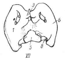
Rice. 23. Axis (epistrophy) of a cow (1), sheep (II), goat (III), horse (IV), pig (V), dog (VI)
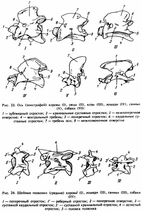
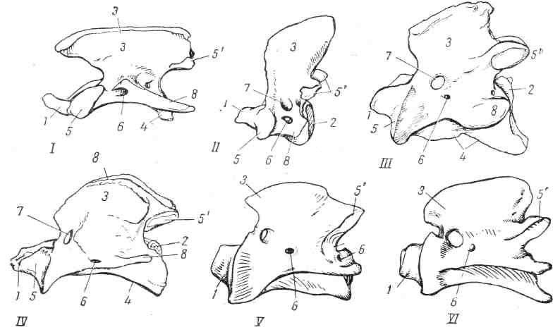
Rice. 24. Cervical vertebrae (middle) cow* (O, horses (II), pigs (III), dogs (IV)
In cattle, the axial vertebra (epistrophy) is massive. The odontoid process is lamellar, semi-cylindrical. The crest of the axial vertebra is thickened along the dorsal margin, and the caudal articular processes protrude independently at its base.
In horses, the axial vertebra is long, the odontoid process is wide, flattened, the crest of the axial vertebra bifurcates in the caudal part, and the articular surfaces of the caudal articular processes lie on the ventral side of this bifurcation.
In pigs, the epistrophy is short, the odontoid process in the form of a wedge has a conical shape, the crest is high (raises in the caudal part).
In dogs, the axial vertebra is long, with a long wedge-shaped odontoid process, the ridge is large, lamellar, protrudes forward and hangs over the odontoid process.
Typical cervical vertebrae - vertebrae cervicales - third, fourth and fifth (Fig. 24).
In cattle, typical cervical vertebrae are shorter than in horses, and the fossa and head are well defined. In the bifurcated transverse process, its cranioventral part (costal process) is large, lamellar, drawn down, the caudodorsal branch is directed laterally. The spinous processes are rounded, well-defined and directed cranially.
Horses have long vertebrae with a well-defined head, vertebral fossa, and ventral crest. The transverse process is bifurcated sagittal plane, both parts of the process are approximately equal in size. There are no spinous processes (scallops in their place).
The upper vertebrae are short, the head and fossa are flat. The costal processes from below are wide, oval-rounded, drawn down, and the caudodorsal plate is directed laterally. There are spinous processes. Very characteristic of the cervical vertebrae of pigs is an additional cranial intervertebral foramen.
In dogs, typical cervical vertebrae are longer than in pigs, but the head and fossa are also flat. The plates of the transverse costal process are almost identical and bifurcate along one sagittal plane (as in a horse). Instead of spinous processes, there are low scallops.
Sixth and seventh cervical vertebrae.
In cattle, on the sixth cervical vertebra, the ventrally strong plate of the costal process is drawn out in a square shape, on the body of the seventh there is a pair of caudal costal facets, the transverse process is not bifurcated. The lamellar spinous process is high. There is no transverse opening, like a horse and a pig.
In horses, the sixth vertebra has three small plates on the transverse process, the seventh is massive, has no transverse foramen, resembles the first thoracic vertebra of a horse in shape, but has only one pair of caudal costal facets and a low spinous process on the body.
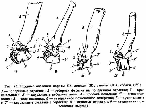
Rice. 25. Thoracic vertebrae of cow (I), horse (II), pig (III), dog (IV)
In pigs, the sixth vertebra has a broad, powerful plate of the transverse process of an oval shape drawn ventrally; on the seventh, the intervertebral foramina are double and the spinous process is high, lamellar, set vertically.
In dogs, the sixth vertebra has a wide plate of the costal process beveled from front to back and downwards; on the seventh, the spinous process is set perpendicularly, has a styloid shape, and the caudal costal facets may be absent.
Thoracic vertebrae - vertebrae thoracicae (Fig. 25).
Cattle have 13 vertebrae. In the region of the withers, the spinous processes are wide, lamellar, caudally inclined. Instead of a caudal vertebral notch, there may be an intervertebral foramen. The diaphragmatic vertebra is the 13th with a steep spinous process.
Horses have 18-19 vertebrae. In the region of the withers, the 3rd, 4th and 5th spinous processes have club-shaped thickenings. The articular processes (except for the 1st) have the appearance of small contiguous articular surfaces. The diaphragmatic vertebra is the 15th (sometimes the 14th or 16th).
Pigs have 14-15 vertebrae, maybe 16. The spinous processes are wide, lamellar, vertically set. At the base of the transverse processes, there are lateral foramens that run from top to bottom (dorsoventrally). There are no ventral ridges. Diaphragmatic vertebra - 11th.
Dogs have 13 vertebrae, rarely 12. The spinous processes at the base of the withers are curved and directed caudally. The first spinous process is the highest; on the latter, ventrally from the caudal articular processes, there are accessory and mastoid processes. Diaphragmatic vertebra - 11th.
Lumbar vertebrae - vertebrae lumbales (Fig. 26).
Cattle have 6 vertebrae. They have a long, slightly narrowed body in the middle part. ventral crest. The transverse costal (transverse) processes are dorsally (horizontally) located, long, lamellar, with pointed jagged edges and ends bent to the cranial side. The articular processes are powerful, widely spaced, with strongly concave or convex articular surfaces.
Horses have 6 vertebrae. Their bodies are shorter than in cattle, the transverse costal processes are thickened, especially the last two or three, on which flat articular surfaces are located along the cranial and caudal edges (in old horses they often synostose). The caudal surface of the transverse costal process of the sixth vertebra is articulated with the cranial margin of the sacral wing. Normally, there is never synostosis here. The articular processes are triangular in shape, less powerful, more closely spaced, with flatter articular surfaces.
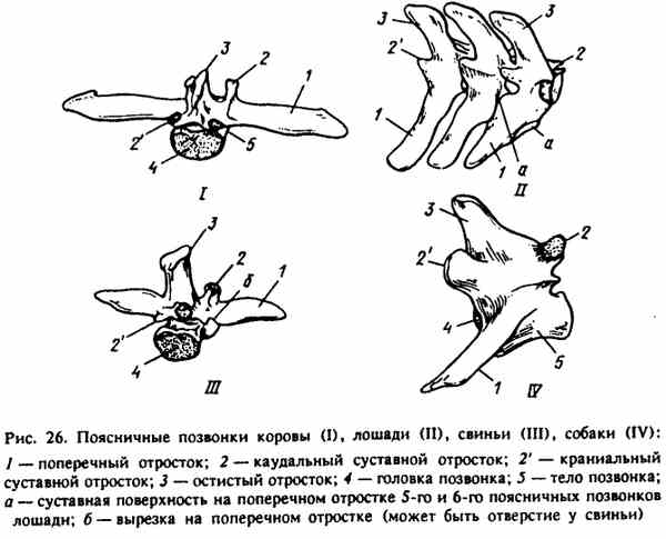
Rice. 26. Lumbar vertebrae of cow (I), horse (I), pig (III), dog (IV)
Pigs have 7, sometimes 6-8 vertebrae. The bodies are long. The transverse costal processes are horizontally arranged, lamellar, slightly curved, have lateral notches at the base of the caudal margin, and lateral foramina closer to the sacrum. The articular processes, like those of ruminants, are powerful, widely spaced, strongly concave or convex, but, unlike ruminants, they have mastoid processes that make them more massive.
Dogs have 7 vertebrae. The transverse costal processes are lamellar, directed cranioventrally. Articular processes have flat articular, slightly inclined surfaces. The accessory and mastoid (on the cranial) processes are strongly pronounced on the articular processes.
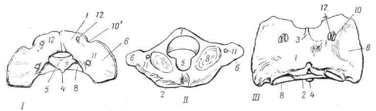
The sacrum - os sacrum (Fig. 27).
In cattle, 5 vertebrae have fused. They have massive quadrangular wings, located almost on a horizontal plane, with a slightly raised cranial margin. The spinous processes are fused, forming a powerful dorsal crest with a thickened edge. The ventral (or pelvic) sacral openings are extensive. Complete synostosis of the vertebral bodies and arches normally occurs by 3-3.5 years.
In horses, 5 fused vertebrae have horizontally arranged triangular wings With two articular surfaces - ear-shaped, dorsal for connection with the wing ilium pelvis and cranial for connection with the transverse costal process of the sixth lumbar vertebra. The spinous processes grow together only at the base.
Pigs have 4 vertebrae fused. The wings are rounded, set on the sagittal plane, the articular (ear-shaped) surface is on their lateral side. There are no spinous processes. Inter-arc holes are visible between the arcs. Normally, synostosis occurs by 1.5-2 years.
In dogs, 3 vertebrae are fused. The wings are rounded, set, as in a pig, in the sagittal plane with a laterally located articular surface. At the 2nd and 3rd vertebrae, the spinous processes are fused. Synostosis is normal by 6-8 months.
Tail vertebrae - vertebrae caudales s. coccygeae (Fig. 28),
Cattle have 18-20 vertebrae. Long, on the dorsal side of the first vertebrae, rudiments of arches are visible, and on the ventral (on the first 9-10) paired hemal processes, which on the 3rd-5th vertebrae can form hemal arches. "The transverse processes are wide, lamellar, ventrally curved.
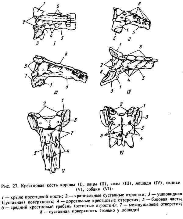
Figure 27. The sacral bone of a cow (1), sheep (I), goat (III), horse (IV), pig (V), dog (VI)
Horses have 18-20 vertebrae. They are short, massive, retain arches without spinous processes, only on the first three vertebrae are the transverse processes flat and wide, disappearing on the last vertebrae.
Pigs have 20-23 vertebrae. They are long, arcuate with spinous processes, inclined caudally, preserved on the first five or six vertebrae, which are flatter, then become cylindrical. The transverse processes are wide.
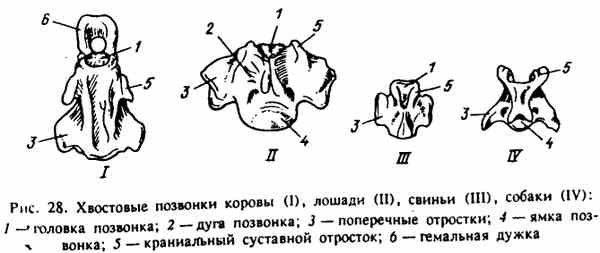
Rice. 28. Tail vertebrae of cow (I), horse (II), pig (III), dog (IV)
Dogs have 20-23 vertebrae. On the first five or six vertebrae, arches, cranial and caudal articular processes are preserved. The transverse processes are large, long, drawn caudoventrally.
Ribs - costae (Fig. 29, 30).
Cattle have 13 pairs of ribs. They have a long neck. The first ribs are the most powerful and the shortest and straightest. Medium lamellar, widening downwards considerably. They have a thinner caudal margin. The posterior ones are more convex, curved, with the head and tubercle of the ribs closer together. The last rib is short, thinning downwards, and may be hanging. It is palpable in the upper third of the costal arch.
Synostosis of the head and tubercle of the rib with the body in young animals does not occur simultaneously and goes from front to back. The head and tubercle of the first rib are the first to fuse with the body. The articular surface of the tubercle is saddle-shaped. The sternal ends of the ribs (from the 2nd to the 10th) have articular surfaces for connection with the costal cartilages, which have articular surfaces at both ends. Sternal ribs 8 pairs.
Horses have 18-19 pairs of ribs. Most of them are of uniform size along the entire length, the first ventrally is significantly expanded, up to the tenth the curvature and length of the ribs increase, then begin to decrease. The widest and lamellar first 6-7 ribs. Unlike ruminants, their caudal margins are thicker and their necks are shorter. The tenth rib is almost four-sided. Sternal ribs 8 pairs.
Pigs often have 14, maybe 12 and up to 17 pairs of ribs. They are narrow, from the first to the third or fourth, the width increases slightly. They have articular surfaces for connection with costal cartilages. In adults, the sternal ends are narrowed; in piglets, they are slightly expanded. Rib tubercles have small flat statutory facets, rib bodies have an indistinct spiral turn. Sternal ribs 7 (6 or 8) pairs.
Dogs have 13 pairs of ribs. They are arched, especially in the middle part. Their length increases to the seventh rib, width - to the third or fourth, and curvature - to the eighth rib. Facet ribs on tubercles convex, sternal ribs 9 pairs.
Breast bone - sternum (Fig. 31).
In cattle, it is powerful, flat. The handle is rounded, raised, does not protrude beyond the first ribs, is connected to the body by a joint. The body expands caudally. On the xiphoid process there is a significant plate of xiphoid cartilage. Along the edges of 7 pairs of articular costal fossae.
In horses, it is laterally compressed. It has a significant cartilaginous addition on the ventral edge, forming a ventral ridge, which protrudes on the handle, rounding off, and is called a falcon. In adult animals, the handle fuses with the body. Cartilage without xiphoid process. Along the dorsal edge of the sternum there are 8 pairs of articular costal fossae.
Rice. 29. Cow ribs (I), horse (II)
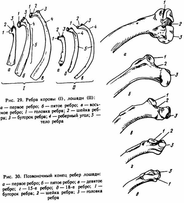
Rice. 30. Vertebral end of horse ribs
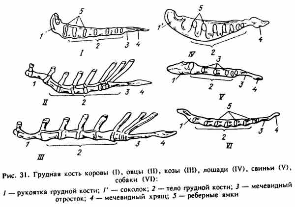
Rice. 31. Breast bone of a cow (I). sheep (II), goats (III), horses (IV), pigs (V), dogs (VI)
In pigs, as in cattle, it is flat, connected to the handle with a joint. The handle, unlike ruminants, in the form of a rounded wedge protrudes ahead of the first pairs of ribs. The xiphoid cartilage is elongated. On the sides b (7-8) pairs of articular costal fossae.
In dogs, it is in the form of a round, well-shaped stick. The handle protrudes in front of the first ribs with a small tubercle. The xiphoid cartilage is rounded, on the sides there are 9 pairs of articular costal fossae.
Thorax - thorax.
In cattle, it is very voluminous, laterally compressed in the anterior part, has a triangular exit. Behind the shoulder blades it greatly expands caudally.
In horses, it is in the form of a cone, long, slightly compressed from the sides, especially in the area of attachment of the shoulder girdle.
In pigs, it is long, laterally compressed, height and width vary in different breeds.
In dogs of a cone-shaped shape with steep sides, the inlet is rounded, the intercostal spaces - spatia intercostalia are large and wide.
Questions for self-examination
1. What is the significance of the apparatus of movement in the life of the organism?
2. What functions does the skeleton perform in the body in mammals and birds?
3. What stages of development in phylo- and ontogenesis does the internal and external skeleton of vertebrates go through?
4. What changes occur in the bones with an increase in static load (with limited motor activity)?
5. How is a bone built as an organ and what are the differences in its structure in young growing organisms?
6. What departments is the vertebral column divided into in terrestrial vertebrates and how many vertebrae are in each department in mammals?
7. Which department axial skeleton Is there a complete bone segment?
8. What are the main parts of the vertebra and what parts are located on each part?
9. In what parts of the spinal column did the vertebrae undergo reduction?
10. By what signs will you distinguish the vertebrae of each department of the spinal column and by what signs will you determine the specific features of the vertebrae of each department?
11. What characteristics structures have an atlas and an axial vertebra (epistrophy) in domestic animals? What is the difference between the atlas of pigs and the axial vertebra of ruminants?
12. By what sign can the thoracic vertebrae be distinguished from the rest of the vertebrae of the spinal column?
13. By what signs can the sacrum of cattle, horses, pigs and dogs be distinguished?
14. What are the main features of the structure of a typical cervical vertebra in ruminants, pigs / horses and dogs.
15. What is the most salient feature have lumbar vertebrae? How do they differ in ruminants, pigs, horses and dogs?
Vertebral column: structure, development, specific features
In its development, the spinal column (columna vertebralis) is formed around spinal cord, forming a bone receptacle for it. In addition to protecting the spinal cord, the spinal column performs other important functions in the body: it is a support for the organs and tissues of the body, supports the head, participates in the formation of the walls of the chest, abdominal cavity and pelvis.
vertebral column(columna vertebralis) consists of separate elements - vertebrae (vertebra). Each vertebra has: a body (corpus vertebrae), a head (caput vertebrae), a fossa (fossa vertebrae), a ventral crest (crista ventralis), an arch (arcus vertebrae), and a vertebral opening (foramen vertebrae) is formed between the arch and the body. All the openings of the vertebrae together form the spinal canal (canalis vertebralis) for the spinal cord, and the caudal and cranial vertebral notches (incisures caudalis et cranialis) form the intervertebral foramen (foramen intervertebrale) for the nerves and blood vessels. The cranial and caudal articular processes (processus articularis cranialis et caudalis) protrude along the edges of the arches, which serve to articulate the vertebrae with each other. The spinous process (processus spinosus) protrudes - fixing muscles and ligaments.
The vertebral column is divided into cervical, thoracic, lumbar, sacral and caudal regions. Transverse processes (processus transversus) in thoracic region needed for the articulation of the vertebrae with the ribs, and the transverse costal, mastoid and spinous (processus costo-transversarium, mamillaris, spinosus) - for muscle attachment.
The number of vertebrae in each department is different and depends on the species characteristics of the animals. Yes, in cervical region most mammals (except sloth and manatee) have 7 vertebrae. They are divided into: 1st - atlas, 2nd - epistrophy, 3rd, 4th, 5th - typical, 6th, 7th.
· 1st(atlas - atlas), consists of two arches (arcus dorsalis et ventralis), on them, respectively, tubercles (tuberculum dorsale et ventrale). The transverse processes form the wings of the atlas (ala atlantis). Under the wing there is an atlas fossa (fossa atlantis), on the wings there are two pairs of holes for vessels and nerves - the wing (foramen alare) and the intervertebral (foramen intervertebrale), there are cranial and caudal articular fossae (fovea articularis cranialis et caudalis). FEATURES: there are no transverse holes on the atlas of the domestic bull.
· 2nd(axial epistrophy - axis), characterized by the presence of a tooth (dens) instead of the head of the vertebra and a ridge (crista dorsalis) instead of the spinous process, also the transverse process (processus transversus) is single.
· 3rd, 4th, 5th- typical. - their transverse processes fused with the costal, forming - transverse costal (processus costo-transversarium), and the spinous processes are tilted towards the head.
· 6th and 7th vertebrae - differ from the rest in shape and are atypical. 6th - instead of a ventral crest, it has a massive ventral plate (lamina ventralis). 7th - does not have a transverse opening, but has caudal costal fossae (fovea costalis caudalis) on the vertebral body.
In the thoracic spine of cattle and dogs, 13 vertebrae each, in pigs 14-17, in horses 18. The thoracic vertebrae (vertebrae thoracicae), together with the ribs and sternum, form the chest. The vertebrae of this department have caudal and cranial costal fossae (fovea costalis caudalis et cranialis), costal facets on the transverse processes (fovea costalis processus transversalis). The spinous process (processus spinosus) is tilted back towards the tail. The spinous processes of the 2nd to 9th vertebrae form the base of the withers (regio interscapularis). The spinous process of the 13th (12th in a pig, 16th in a horse, and 11th in a dog) vertebra stands vertically - diaphragmatic. On the transverse processes (processus transversus) are mastoid processes (processus mamillaris).
AT lumbar of the spine in cattle and horses, 6 vertebrae each, in pigs and dogs, 7 each. Lumbar vertebrae (vertebrae lumbales), characterized by the presence of long, flat transverse processes and well-developed articular processes. transverse processes with sharp, uneven edges and bent forward towards the head. The spinous processes stand vertically. The cranial articular processes form semi-cylindrical bushings, and the caudal processes form the same blocks.
AT sacral region The vertebrae of the spine (vertebrae sacrales) fuse into one bone - the sacrum (os sacrum), which consists of 5 vertebrae in cattle and horses, 4 in pigs, and 3 in dogs.
The spinous processes have merged into the medial sacral crest (crista sacralis mediana), there are no interannual foramen. Intervertebral notches formed 4 pairs of dorsal and ventral sacral foramina (foramina sacralia dorsalia et ventralia). The transverse processes have merged - jagged lateral parts (partes lateralis). The first two transverse processes formed the wings of the sacrum (ala sacralis). On the wings dorsally there is an ear-shaped cover (facies auricularis), the ventral cover is pelvic (facies pelvina). On the vent. Transverse lines (lineae transversae) are visible again, a vascular trough passes here. The head ventrally forms a sacral promontory (promontorium). There is also a sacral canal (canalis sacralis).
The tail section of the spine is the most variable in terms of the number of vertebrae, which are 20-23 in dogs, 20-25 in pigs, 18-20 in cattle, and 18-20 in horses. In the structure of the caudal vertebrae (vertebrae caudales (coccygeae)) there is a gradual reduction of the arc. On the ventral side, from the 2nd to the 13th, hemal processes (processus hemalis) are well developed.
The cervical region of the skeleton plays the role of a powerful single-arm flexible lever, at the front end of which is the head of the animal.
Despite significant fluctuations in the length of the neck in different animals, the cervical vertebrae - vertebrae cervicales - in mammals, with few exceptions, the same number - seven. In their structure, the 3rd, 4th, 5th and 6th vertebrae are most similar to each other. All of them are massive; have two-branched transverse costal processes and transverse openings - for. transversarium - at their base; powerful articular processes are connected by lateral ridges, which gives the cervical vertebrae, together with their transverse costal processes, the shape of tetrahedral grooved prisms; small spinous processes directed cranially; in addition, these vertebrae in animals with long neck elongated in length and have pronounced heads and fossae; on the contrary, in animals with a short neck (for example, pigs), they are shortened, and the heads and fossae of the vertebrae are flat (Fig. 10).
Peculiarities.
In a dog, the heads and fossae of the vertebrae are flat, set obliquely in relation to the body. The ventral crests protrude only at the caudal ends of the vertebral bodies. The vertebral arches are very long, and the foramina between the vertebrae are small. The spinous process on the 3rd vertebra is absent, while on the rest, the length of the spinous processes increases in the caudal direction. Typical, in the form of tubercles, the mastoid processes are located on the caudal articular processes. The costal processes are directed cranially.
Rice. 10. Middle cervical vertebrae:
I - dogs; II - pigs; III - cows; IV - horses. 1 - caput vertebrae (vertebral head); 2 - fossa vertebrae (fossa of the vertebra); 7'-foramen vertebrate laterale (lateral vertebral foramen); 9 - processus articularis cranialis (cranial articular process); 9' - processus articularis caudalis (caudal articular process); 10 - processus spinosus (spinous process); 11 - processus costo-transversarius (transverse process); 12 - processus mamillaris (mastoid process); 13 - processus costo-transversarius (transverse costal process); 13′ - processus costarius (costal process); 17 - foramen transversarium (megatransverse hole).
The pig's vertebrae are massive but very short; heads and pits are flat; the arches are narrow, and the foramina between the arches are wide. The spinous processes are narrow, long. The costal processes are very wide, directed ventrally, and tiled on the processes of the cranially lying vertebrae. At the base of the transverse costal processes, in addition to the transverse foramina, there are dorsoventral foramina. The ventral crest is absent.
In cattle, the vertebrae are massive and short; heads and pits are well expressed; the spinous processes are developed, have thickened ends, their length increases in the caudal direction. The costal processes are located ventrally from the transverse processes and are deflected anteriorly. On the 6th vertebra, the costal process is wide and long; the ventral crest is absent.
The horse's vertebrae are very massive and long; their heads and fossae are strongly pronounced; spinous processes are replaced by roughness. The transverse costal processes are provided with two branches; one of them (costal process) is directed cranially, and the other (transverse process) is directed caudally. The ventral crests are strongly pronounced, except for the crest of the 6th vertebra. The length of the vertebrae decreases from the 3rd to the 6th; on the 6th vertebra, the transverse foramen is the widest, and the costal process is also wide.
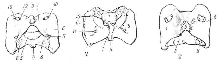
Rice. 11. Atlas: I-dogs; II - pigs, from the caudal side; III - cows; IV - horses, from the dorsal surface; V-. horses, from the ventral surface; VI - sheep; VII-. goats. 1 - ar-cus dorsalis (dorsal arch); 2 - arcus ventralis (ventral arch); 3 - tuberculum dorsale (dorsal tubercle); 4 - tuberculum ventrale (ventral tubercle); 5 - facies articularis dentis (articular surface for the odontoid process); 6 - ala atlantis (wing of the Atlantean); 7 - fovea articularis cranialis (cranial articular fossa); 8 -facies articularis caudalis (caudal articular surface); 9 - fossa atlantis (wing fossa of the atlas); 10 - foramen alare (wing hole); 10′ - incisura alaris (wing notch); 11- foramen transversarium (intertransverse opening); 12 - foramen intervertebrale (intervertebral foramen).
Among the cervical vertebrae, the first two and the last differ significantly in their development from each other and from all the others.
The first cervical vertebra, the atlas, provides greater head mobility. Its appearance is ring-shaped with lateral processes - the wings of the atlas (Fig. 11). On the Atlanta, the dorsal and ventral arches are distinguished - arcus dorsalis et ventralis. The spinous process is replaced by a dorsal tubercle - tuberculum dorsale. The ventral arch corresponds to the body of the atlas; from the side of the vertebral foramen, it bears a facet for the odontoid process of the epistrophy - facies articularis dentis, and from the ventral side - the ventral tubercle - tuberculum ventrale, developed more strongly than the dorsal one. The transverse processes are fused with the articular ones, as a result of which they are very powerful and are called the wings of the atlas - ala atlantis. The bases of the wings form articular fossae from the cranial end - fovea articularis cranialis - for connection with the condyles occipital bone, and at the caudal end they protrude convex articular surfaces - i acies articularis caudalis - for connection with the 2nd cervical vertebra. On the ventral surface of the wings are wing pits - fossa atlantis. The anterior end of the wing pierces the wing opening - foramen alare, leading to the wing fossa. The alar opening is connected by a groove to the intervertebral foramen - foramen intervertebrale, and the latter - to the spinal canal.
Peculiarities. In the dog, the wings of the atlas are flat, thin, set almost horizontally, extended latero-caudally. Wing pits are small.
Rice. 12. Epistropheus (epistropheus):
I - dogs; II - pigs; III - cows; IV - horses; V - sheep; VI - goats. 1 - dens epistrophei (dentate process); d - fossa vertebrae (fossa of the vertebra); 3 - crista epistrophei (comb of epistrophy); 4 - crista ventralis (ventral crest); 5 - processus articularis cranialis (cranial articular process); 5' - rgos. articularis caudalis (caudal articular process); 6 - for. transversarium (intertransverse opening); 7-for. intervertebrale (intervertebral foramen); 8 - rgos. transversus (transverse process).
Instead of a wing hole, there is a wing notch - incisura alaris. Dorsal arch wide, without tubercle. The ventral arch is narrow, completely covered with a facet for the odontoid process of the epistrophy. The transverse opening - foramen transversarium - leads to the dorsal surface of the wing into the wing fossa.
In the pig, the wings of the atlas have well-defined anterior and posterior angles and an arcuate margin. At the base of the wings, an intertransverse canal begins behind - canalis transversarius, which goes into a flat wing fossa. The ventral arch is already dorsal and has a deep notch for the odontoid process of the epistrophy. The ventral tubercle is large and directed caudally. The dorsal tubercle is well developed.
In cattle, the wings of the atlas form cranial and more widely spaced caudal angles. Wing fossa small. The facet for the odontoid process of the epistrophy occupies only the caudal half of the arch. There is no transverse opening. For sheep and goats, see fig. eleven.
In the horse, the wings of the atlas with rounded edges are strongly bent ventrally, as a result of which the wing fossae are deep. There is a transverse opening - foramen transversarium. The facet for the odontoid process of the epistrophy occupies only the caudal half of the ventral arch.
The second cervical vertebra is the axis, or epistrophy - axis, s. epistropheus (Fig. 12) - characterized by a significant odontoid process - dens epistrophei, corresponding to the head of the vertebra, and a spinous process in the form of an epistrophe ridge - crista epistrophei, weak (not branching) transverse costal processes with transverse foramens - foramen transversarium - at the base. An intervertebral foramen opens in front of the transverse process. The cranial articular processes are very powerful and are located behind and laterally from the odontoid process.
Peculiarities. In a dog, the odontoid process is cylindrical; the crest of the epistrophy hangs cranially over the odontoid process, and merges caudally with the caudal articular processes. The intervertebral foramen was replaced by a notch.
In a pig, the odontoid process is blunt, cone-shaped. The crest of the epistrophy is pterygoid, narrow, high; its posterior margin is raised dorsally. The vertebra as a whole is massive, but short.
In cattle, the odontoid process looks like a hollow half-cylinder, and the crest of the epistrophy is a square plate. For sheep and goats, see fig. 12.
The horse has an odontoid process with a flat dorsal surface and a convex (articular) ventral surface. A very powerful crest of the epistrophy bifurcates caudally and fuses with the caudal articular processes. The ventral crest, crista ventralis, is quite well defined. The epistropheus as a whole is very massive and the longest of all the vertebrae.
The 7th cervical vertebra is provided with only one pair of costal facets - foveae costales caudales - at the caudal end of the body. The transverse process does not branch, and there is no transverse foramen at its base. The spinous process is more powerful than on the other cervical vertebrae.
Peculiarities. In the dog, the head and fossa of the vertebrae are flat; the spinous process is psiloid and set perpendicular to the vertebral body. Rib facets may be absent.
In a pig, the head and fossa are flat; the intervertebral foramina are double, as on all vertebrae in general.
In cattle, the head and fossa of the vertebrae are large; ventral crest absent; the spinous process is high and wide.
The horse has a strongly developed head and fossa of the vertebrae and a weakly developed spinous process and ventral crest.