Modern scientists of anatomy. Research methods. Questions for self-control
Outstanding Russian scientists - anatomists
The first stages of the development of anatomy and medicine of the Russian state in the 18th century were illuminated by the genius of Peter I, who showed interest in training doctors in Holland, where he attended lectures and anatomical theaters of professors F. Ruysch, G. Burgave and A. Van Leeuwenhoek. To educate Russians, Peter the Great acquired an anatomical collection for the Kunstkamera, which, by his decree, has been constantly replenished since 1718 with embryological and teratological preparations, which have been preserved in St. Petersburg to this day. Upon returning from abroad to Moscow, the tsar organized a series of lectures and dissections for the boyars, and studied at the Moscow Anatomical Theater to dissect corpses and perform surgical operations. Subsequently, such events became regular and were carried out in hospitals, a medical school organized by Peter at the Academy of Sciences.
He also gained fame as a botanist and was director of the Botanical Garden of Padua. Miguel Servetus, a Spanish physician and theologian who was executed because of his beliefs by the Calvinist dictatorship of Geneva. He studied law at the University of Toulouse, medicine at the University of Paris and Montpellier, and theology at Leuven. Even though he was considered a Catholic, he supported a personal concept of the dogma of the Holy Trinity and asked permission to visit the theocratic city created by Calvin in Geneva, where he was arrested while attending a mass.
In Moscow, St. Petersburg, Barnaul, Kronstadt and other cities (more than 30), medical schools were opened at hospitals, in which doctors were initially trained by foreign anatomists and surgeons: N.L. Bidloo, A. De-Tils, L.L. Blumentrost and others. D. Bernoulli, I. Weitbrecht, I. Duvernois, and subsequently the great M.V. contributed to the formation of anatomy and physiology at the Petrine Academy of Sciences. Lomonosov is a candidate of medicine at the University of Magdeburg.
Accused of heresy and blasphemy against Christianity, he died, burned at the stake. The religious views of the Servetians were guided by the Catholics and Protestants of the time. In the error of the Trinity, he rejected the tripartite personality of God and the ritual of baptism. His scientific contributions were also notable: A Restoration of Christianity, published shortly before his death, contains the first rigorous description of the pulmonary circulatory system.
William Harvey, an English physician who discovered the circulation of blood and the role of the heart in its movement, thereby refuting Galen's theories and laying the foundations of modern physiology. He went to Padua, Italy, where he studied for five years with the famous anatomist Fabrizio, who was already studying venous valves.
A student and follower of Academician M.V. Lomonosov was A.P. Protasov, who also became an academician, teaching a university course in anatomy. He is known for his work on the anatomical and physiological structure of the stomach, the compilation of an anatomical dictionary in Russian, and forensic medical autopsies of corpses.
K.I. Shchepin- one of the first Russian anatomist professors, taught anatomy, physiology and surgery in Russian. At the St. Petersburg and Moscow hospital schools he created programs for these disciplines and introduced a clinical focus into them. In his lectures he used data from microscopic anatomy for the first time. He died in Kyiv during the elimination of the plague epidemic.
He came to these conclusions not only through a long series of dissections, but also through studies of the movement of the heart and blood in a wide variety of living animals. The accuracy of his observations established a model for future biological research. In this transcendental work he explained the experimental method and offered an accurate description of the mechanism of the circulatory system. Since he did not have a microscope, the only important part of the process that he missed was the role of capillaries. However, he postulated his existence, which was soon confirmed by the Italian Marcelo Malpighi.
M.I. Shein- translated from German language anatomy textbook by Ludwig Geister, which was published for the first time in 1757 in St. Petersburg. At the same time, he believed that correct knowledge of the human structure is useful for health, healing, and treatment. He introduced new anatomical terms in Russian, which have survived to this day, and created the first Russian anatomical atlas.
Questions for self-control
De Motu Cordiz caused Harvey to come under harsh criticism from some of his contemporaries, although this was largely offset by subsequent recognition of the value of his contributions. Marcello Malpighi, Italian physiologist whose discoveries in microscopic anatomy banished ancient medical beliefs and opened the way to modern physiology and histology.
As professor of medical theory at the University of Pisa, he began his microscopic observations and took a very serious critical attitude towards the assumptions on which physiology and medicine were based. Until then, the prevailing belief was that blood was transformed into flesh on the periphery of the body.
N.M. Maksimovic-Ambodik– professor of midwifery (obstetric) sciences, prepared the first Russian anatomical nomenclature and wrote the “Anatomical and Physiological Dictionary”. Modern names of organs did not appear immediately, for example, the pancreas was called “all-meat”, “tongue-shaped”, the artery was called the vein, the vein was called the vein. That is why the work on the selection of scientific anatomical names, carried out over a whole century, was so important.
He was the first to discover red blood cells and showed that they were the ones that gave him his color. He also identified taste buds and described the chicken embryo, silkworm development, and plant structure. Malpighi believed that living matter consists of tiny glands that separate or mix physical fluids. Although he misinterpreted the microscopic functions of organs, he paved the way for cell theory and histology.
His discoveries about the occult functioning of organisms forced doctors of the time to seriously reconsider firmly established theories. He was born in Forli and studied at the University of Bologna. He was appointed professor of anatomy at the University of Padua. He reported numerous examinations of corpses with corresponding medical data and discovered the relationship between diseases and physical transformations of organs.
As a result from the first anatomical terminology many Old Slavonic designations have disappeared, such as lyadovia - lower back, ramo - humerus, femoral vein, lucid vein - pulmonary vein, ridge - spine, spinal bone marrow - spinal cord. But many new names were immediately fixed in Russian nomenclature: clavicle, ankle, etc., and some were recognizably modified: tibia - tibia, epigastric region from the old name of the xiphoid process of the sternum - spoon. Thus, the origins of Russian anatomical names were Russian vocabulary and Greco-Latin terminology.
His descriptions of the effects of disease on the heart, lungs and liver were particularly accurate. The history of medicine is long and distinct, as healers have sought to cure diseases and ease wounds since the dawn of mankind. It includes witch doctors and pagan priests who used a mixture of rituals and medical techniques to treat illnesses.
Andres Vesalio and modern human anatomy
One of these important areas was one that showed how medical practice began to resemble our modern systems.
Discovery of bacteria
Anton van Leeuwenhoek is considered the father of microbiology. Van Leeuwenhoek is known for them. Semmelweis and hand washing. Some of the Nobel Prizes are in the field of "Medicine or Physiology". Winner Discovery: Barbara McClintock.P.A. Zagorsky- Academician, when compiling a Russian anatomy textbook, he carefully selected the main Russian terms. He founded an anatomical school in St. Petersburg and studied teratology and comparative anatomy. Prepared a worthy student - Professor I.V. Buyalsky, who published “Anatomical and Surgical Tables”, wrote a textbook with anatomical justification surgical operations, invented many instruments, proposed new methods of embalming. I.B. Buyalsky was engaged in the preservation of anatomical preparations, using solutions of mercuric chloride to inject blood vessels, and its powder was poured into body cavities. Contributed to the development of anatomy at the St. Petersburg school A.M. Shumlyansky, who discovered capsules around the vascular glomeruli of the kidney (nephron capsule), establishing direct connections between arterial capillaries in the vascular glomerulus. Academician K.F. Wolf for a long time he headed the anatomical department of the St. Petersburg Kunstkamera. His teratological collections formed the basis for work on defects and deformities, which gave rise to the development of a new anatomical science - teratology.
II. Physiological methods
Opening of the winners: Erwin Neher and Bert Sackmann. Opening of which Winner: Günter Blobel. Winners: Paul Lauterbur and Sir Peter Mansfield. Winners: Françoise Barré-Sinoussi and Luc Montagnier. In the Middle Ages, the study of human anatomy in Europe was based on the knowledge of Ancient Greece, thanks to Hipparchus and Ancient Rome, thanks to Sels and mainly Galen. These pillars are described in broad strokes of both human anatomy and the physiology of human functioning.
With the opening of Italian universities, everything began to gradually change. The doctors in charge of the teaching began to count with surgeons in their classes to dissect corpses and thus show the insides to their students. The trade of these surgeons was less valuable, and their skill left much to be desired. Everything turned out to be extremely boring for all the students, who preferred to take advantage of the enormous oratorical abilities of the medical teachers.
Professor E.O. Mukhin taught anatomy at Moscow University. After the invasion of Napoleon and the fire in Moscow, the anatomical museum was restored, containing up to 5,000 preparations. In 1812, his textbook “Anatomy Course” was published, in which the author promoted Russian anatomical terminology.
Professor D.N. 3ernov for many years headed the Moscow Department of Anatomy; successfully studied the sense organs, the variability of the furrows, convolutions of the brain and criticized Cesaro Lombroso's theory about the hereditary factors of the criminal personality, about the correspondence of certain types of face and brain to aggressive and malicious behavior.
Topic: “The study of fabrics. Epithelial tissue"
The son of a doctor, his education was oriented toward Renaissance humanism, following the guidelines of the new schools of the time. Meanwhile, in his home, he used ancient anatomy books from his father, developing an interest in dissecting animals. Thanks to Silvius's great ability to reveal, Vesalio was imbued with all the knowledge of Galen. After a few months, Silvius became aware of Vesalius' ability to dissipate, so he offered to replace the surgeon of his classes, which Vesalio accepted without hesitation. Four years later, after completing his studies, his skill had improved markedly, and he used it to move to Padua, where at the age of 22 he was appointed professor of anatomy.
V.A. Betz- a representative of the Kyiv anatomical school, who discovered large pyramidal cells in the convolutions of the brain, named after his last name. Professor in Kharkov A.K. Belousov studied the innervation of blood vessels, proposed a new method of injection of anatomical drugs.
The formation of anatomy as a science and educational subject in the 18th and 19th centuries happened at first thanks to foreign specialists invited by Peter I, who very soon trained students and followers in St. Petersburg and Moscow. Both schools became leading ones; they sent their graduates to provincial universities, who founded departments, established anatomical science, and trained doctors.
It was then that he began to revolutionize the world of medicine through his teaching. Going further than Silvius to use him in his classes, Vesalio suppressed the surgeon from his classes, but decided to take his place, dissecting corpses, explaining to his students that they became more than 500 at a time, your knowledge of anatomy.
Outstanding Russian scientists - anatomists
The greatness of this book lies not in his texts, but in his illustrations, for which he helped his friend Jan Stephen van Calcar, a student of Titian. On the cover of the first edition of this great work we find a presentation of one of the master classes given as a professor of anatomy in Padua. Already exploring our pages, we find illustrations human body with detail they have never been presented with before. A series of plates representing the human skeleton in all types of poses, another series of plates representing the muscles of the human body, together with partial dissections of the same.
The development of healing in the east of Russia was influenced by Tibetan and Chinese medicine, traditional medicine of the peoples of the North and Far East. In Buryatia, from the middle of the 18th century, medical schools (mamba-datsans) appeared at Buddhist monasteries. They use medical literature, diagnostic and treatment methods, and tools from Mongolia, Tibet, India and China for training. Thus, the Atsagat school was founded by Emgi-Lama Ireltuev, a skilled healer and teacher. The full course of study took 6 years, and the human structure was always considered under the leading influence of function. In the middle of the 19th century, the Badmaev brothers Tsultim and Zhamsadin studied Tibetan medicine in the Aginsky datsan (monastery), who later converted to Orthodoxy under the names of Alexander and Peter (godsons of the emperor Alexandra III). They got their university degree medical education in St. Petersburg. Both had extensive practice in the capital in aristocratic and noble circles, and participated in political and palace intrigues.
The poses presented in these first two series reflect the methods used by Vesalio during his classes to demonstrate anatomy to his students. In later sheets there appears a rough description of blood vessels and nerves, always trying to represent their dissections. Then you can see illustrations of all the internal organs of the human body in Galenic order. The senses are not missing in the later pages, where there is a curious dissection of an animal eye that stands in for a human, as well as images of pregnant women and their fetuses.
Other scientists and practitioners also made a certain contribution to the development of oriental medicine in Russia. So the famous Altai geographer and ethnologist G.N. Potanin published an article about Buryat names medicinal plants, used in Tibetan and folk medicine. Kalmyk Dambo Ulyanov, the chief physician and lama of the Don Cossack Army, translated medical treatises “Chzhud-shi”, “Lkhantab” and others from Tibetan into Russian.
To finish the book, there are some sheets that have all the tools used to open them drawn. Artistically, Factory is a unique book that enjoys enormous ambition in its aesthetic. For this reason it was copied in all the great universities of Europe, as well as in the most important courts. Even today they are used as a representation of the human body with great devotion, despite the passage of more than four centuries. But if we moved to more medical institution Vesalio did not dare to explain anything himself, resorting to Galen to talk about the physiognomy of the human body.
In Siberia, the first university was opened in the city of Tomsk in 1870. The Faculty of Medicine opened there in 1876 under the leadership of famous professors of the Kazan school A.S. Dogel and A.E. Smirnova. All subsequent Siberian and Far Eastern medical institutes and faculties were opened during the years of Soviet power. The Altai Medical Institute appeared in Barnaul in 1954 in connection with the massive development of virgin and fallow lands. His professional development took place under the influence and direct participation of scientists and teachers of the capital and Tomsk medical universities, but in a certain sense he became the legal successor of the Barnaul medical school of the 18th century, opened at the behest of Peter I.
Of course, at the time of writing, he left all his training aside to describe only what he perceived with his own eyes in the showdowns he performed. History of Anatomy Anatomy is like a lady who is hard to defeat, but once defeated, she is never forgotten. Developmental anatomy: studies the changes that occur from the fertilized egg to the adult state. Doctor Radiographic Anatomy: studies body structures using x-rays. Dr. Systemic anatomy: studies specific body systems, such as the cardiovascular system or the Nervous Doctor system The dispersal of human cadavers has provided the basis for understanding the structure and function of the human body for many centuries. Hippocrates attributed diseases to more natural causes than the displeasure of the gods. He named the Aorta and contrasted arteries and veins. Alexandria had medical schools and a large library. He performed vivisections and dissection of human corpses. He described the skull, the eye, and various visceral organs. He described the meninges, the brain, the cerebellum. He described the sensory and motor nerve. He isolated the cerebellum and determined that the brain was the source of all nerves, classifying them into motor and sensory. At the beginning of the 3rd century BC, two surgeons in Alexandria, Herophilus and Erasistratus, conducted the first scientific studies aimed at identifying the functioning of human anatomy. At present, the value of his contribution to science would be considered very high. But Celsus, the Roman writer of medical history, vigorously justified the suffering of these criminals as providing a means for innocent people of future generations. His anatomy was based on animal dissection rather than human cadavers. He correctly described many brain structures, the structural differences between veins and arteries, and the valves of the heart. The anatomists of that time were conditioned to accept the outdated information of Aristotle and Galen, and if in any dissection they found something different from previous teachings, anatomists dr. Leonardo da Vinci made hundreds of anatomical drawings since the dissection, but unfortunately he had little influence on the anatomists of his time. Structural anatomy: studies the cellular level, tissues and organs. . The basis of medicine is anatomy.
Research methods
Brief history anatomy development
And physiology
The development and formation of ideas about anatomy and physiology begin in ancient times.
Among the first anatomists known to history are Alkemona from Cratona, who lived in the 5th century. BC e. He was the first to dissect (dissect) the corpses of animals in order to study the structure of their bodies, and suggested that the sense organs communicate directly with the brain, and the perception of feelings depends on the brain.
Hippocrates(c. 460 - c. 370 BC) - one of the outstanding medical scientists Ancient Greece. He attached paramount importance to the study of anatomy, embryology and physiology, considering them the basis of all medicine. He collected and systematized observations about the structure of the human body, described the bones of the roof of the skull and the connections of bones with sutures, the structure of the vertebrae, ribs, internal organs, the organ of vision, muscles, and large vessels.
The outstanding natural scientists of their time were Plato (427-347 BC) and Aristotle (384-322 BC). Studying anatomy and embryology, Plato discovered that the brain of vertebrates develops in the anterior regions spinal cord. Aristotle, opening the corpses of animals, he described their internal organs, tendons, nerves, bones and cartilage. In his opinion, the main organ in the body is the heart. He named the largest blood vessel the aorta.
Great influence on development medical science and anatomy had Alexandria School of Physicians, which was created in the 3rd century. BC e. Doctors of this school were allowed to dissect human corpses for scientific purposes. During this period, the names of two outstanding anatomists became known: Herophilus (b. c. 300 BC) and Erasistratus (c. 300 - c. 240 BC). Herophilus described the meninges and venous sinuses, cerebral ventricles and choroid plexuses, optic nerve and eyeball, duodenum and vessels of the mesentery, prostate. Erasistratus described the liver, bile ducts, heart and its valves quite fully for his time; knew that blood from the lung enters the left atrium, then into the left ventricle of the heart, and from there through the arteries to the organs. The Alexandrian school of medicine also discovered a method for ligating blood vessels during bleeding.
The most outstanding scientist in various fields of medicine after Hippocrates was the Roman anatomist and physiologist Claudius Galen(approx. 130 - approx. 201). He first began teaching a course in human anatomy, accompanied by dissections of animal corpses, mainly monkeys. Dissection of human corpses was prohibited at that time, as a result of which Galen, facts without due reservations, transferred the structure of the animal's body to humans. Possessing encyclopedic knowledge, he described 7 pairs (out of 12) of cranial nerves, connective tissue, muscle nerves, blood vessels of the liver, kidneys and other internal organs, periosteum, ligaments.
Important information was obtained by Galen about the structure of the brain. Galen considered it the center of sensitivity of the body and the cause of voluntary movements. In the book “On the Parts of the Human Body,” he expressed his anatomical views and considered anatomical structures in inextricable connection with function.
A Tajik doctor and philosopher made a great contribution to the development of medical science Abu Ali Ibn Son, or Avicenna(c. 980-1037). He wrote the “Canon of Medical Science,” which systematized and supplemented information on anatomy and physiology, borrowed from the books of Aristotle and Galen. Avicenna's books have been translated into Latin and was reprinted more than 30 times.
Since the XVI-XVIII centuries. In many countries, universities were opened, medical faculties were established, and the foundation of scientific anatomy and physiology was laid. An especially great contribution to the development of anatomy was made by the Italian scientist and artist of the Renaissance. Leonardo da Vinci(1452-1519). He anatomized 30 corpses, made many drawings of bones, muscles, and internal organs, providing them with written explanations. Leonardo da Vinci laid the foundation for plastic anatomy.
A professor at the University of Padua is considered the founder of scientific anatomy. Andras Vesalius(1514-1564), who, based on his own observations made during autopsies of corpses, wrote a classic work in 7 books “On the structure of the human body” (Basel, 1543). In them he systematized the skeleton, ligaments, muscles, blood vessels, nerves, internal organs, brain and sense organs. Vesalius's research and the publication of his books contributed to the development of anatomy. Subsequently, his students and followers in the 16th-17th centuries. made many discoveries and described in detail many human organs. The names of some organs of the human body are associated with the names of these scientists in anatomy: G. Fallopius (1523-1562) - fallopian tubes; B. Eustachius (1510-1574) - Eustachian tube; M. Malpighi (1628-1694) - Malpighian corpuscles in the spleen and kidneys.
Discoveries in anatomy served as the basis for deeper research in the field of physiology. The Spanish physician Miguel Servetus (1511-1553), a student of Vesalius R. Colombo (1516-1559), suggested that blood passes from the right half of the heart to the left through the pulmonary vessels. After numerous studies, the English scientist William Harvey(1578-1657) published the book “An Anatomical Study on the Movement of the Heart and Blood in Animals” (1628), where he provided evidence of the movement of blood through the vessels of the systemic circulation, and also noted the presence of small vessels (capillaries) between arteries and veins. These vessels were discovered later, in 1661, by the founder of microscopic anatomy, M. Malpighi.
In addition, W. Harvey introduced vivisection into the practice of scientific research, which made it possible to observe the functioning of animal organs using tissue sections. The discovery of the doctrine of blood circulation is considered to be the founding date of animal physiology.
Simultaneously with the discovery of W. Harvey, a work was published Casparo Azelli(1591-1626), in which he made an anatomical description lymphatic vessels mesentery of the small intestine.
During the XVII-XVIII centuries. not only new discoveries in the field of anatomy appear, but a number of new disciplines begin to emerge: histology, embryology, and somewhat later - comparative and topographic anatomy, anthropology.
For the development of evolutionary morphology, the teaching played an important role C. Darwin(1809-1882) about the influence of external factors on the development of forms and structures of organisms, as well as on the heredity of their offspring.
Cell theory T. Schwann (1810-1882), evolutionary theory Ch. Darwin set a number of new tasks for anatomical science: not only to describe, but also to explain the structure of the human body, its features, to reveal the phylogenetic past in anatomical structures, to explain how his individual characteristics developed in the process of historical development of man.
To the most significant achievements of the 17th-18th centuries. refers to what was formulated by the French philosopher and physiologist Rene Descartes the idea of “reflected activity of the body.” He introduced the concept of reflex into physiology. Descartes' discovery served as the basis for further development physiology on a materialistic basis. Later ideas about the nervous reflex, reflex arc, meaning nervous system in the relationship between the external environment and the body were developed in the works of the famous Czech anatomist and physiologist G. Prohaski(1748-1820). Advances in physics and chemistry have made it possible to apply more precise methods research.
In the XVIII-XIX centuries. Particularly significant contributions to the field of anatomy and physiology were made by a number of Russian scientists. M. V. Lomonosov(1711-1765) discovered the law of conservation of matter and energy, expressed the idea of the formation of heat in the body itself, formulated a three-component theory of color vision, and gave the first classification of taste sensations. Student of M. V. Lomonosov A. P. Protasov(1724-1796) - author of many works on the study of the human physique, structure and functions of the stomach.
Professor of Moscow University S. G. Zabelin(1735-1802) lectured on anatomy and published the book “A Tale on the Structures of the Human Body and How to Protect Them from Diseases,” where he expressed the idea of the common origin of animals and humans.
In 1783 I. M. Ambodik-Maksimovich(1744-1812) published the “Anatomical and Physiological Dictionary” in Russian, Latin and French, and in 1788 A. M. Shumlyansky(1748-1795) described the capsule in his book glomerulus and urinary tubules.
A significant place in the development of anatomy belongs to E. O. Mukhina(1766-1850), who taught anatomy for many years, wrote training manual"Anatomy Course".
The founder of topographic anatomy is N. I. Pirogov(1810-1881). He developed an original method for studying the human body using cuts from frozen corpses. Author of such famous books as “A Complete Course in Applied Anatomy of the Human Body” and “ Topographic anatomy, illustrated by cuts made through the frozen human body in three directions." N.I. Pirogov especially carefully studied and described the fascia, their relationship with blood vessels, giving them great practical importance. He summarized his research in the book “ Surgical anatomy arterial trunks and fascia."
Functional anatomy was founded by an anatomist P. F. Les-gaft(1837-1909). Its provisions on the possibility of changing the structure of the human body through exposure physical exercise on the functions of the body form the basis of the theory and practice of physical education. .
P. F. Lesgaft was one of the first to use the radiography method for anatomical studies, the experimental method on animals and methods of mathematical analysis.
The works of famous Russian scientists K. F. Wolf, K. M. Baer and X. I. Pander were devoted to the issues of embryology.
In the 20th century functional and experimental directions in anatomy were successfully developed by such research scientists as V. N. Tonkov (1872-1954), B. A. Dolgo-Saburov (1890-1960), V. N. Shevkunenko (1872-1952), V. . P. Vorobyov (1876-1937), D.A. Zhdanov (1908-1971) and others.
The formation of physiology as an independent science in the 20th century. significantly contributed to advances in the field of physics and chemistry, which gave researchers precise methodological techniques that made it possible to characterize the physical and chemical essence of physiological processes.
I. M. Sechenov(1829-1905) entered the history of science as the first experimental researcher of a complex phenomenon in the field of nature - consciousness. In addition, he was the first who managed to study gases dissolved in the blood, establish the relative effectiveness of the influence of various ions on physical and chemical processes in a living organism, and clarify the phenomenon of summation in the central nervous system (CNS). I.M. Sechenov gained the greatest fame after the discovery of the process of inhibition in the central nervous system. After the publication of I.M. Sechenov’s work “Reflexes of the Brain” in 1863, the concept of mental activity was introduced into the physiological foundations. Thus it was formed new look on the unity of the physical and mental foundations of man.
The development of physiology was greatly influenced by the work I. P. Pavlova(1849-1936). He created the doctrine of the higher nervous activity of humans and animals. Studying the regulation and self-regulation of blood circulation, he established the presence of special nerves, some of which strengthen, others delay, and others change the strength of heart contractions without changing their frequency. At the same time, I.P. Pavlov also studied the physiology of digestion. Having developed and applied in practice a number of special surgical techniques, he created new physiology digestion. Studying the dynamics of digestion, he showed its ability to adapt to excitatory secretion when consuming various foods. His book “Lectures on the work of the main digestive glands” became a guide for physiologists around the world. For his work in the field of digestive physiology in 1904, I. P. Pavlov was awarded the Nobel Prize. His discovery of the conditioned reflex allowed him to continue the study of mental processes that underlie the behavior of animals and humans. The results of many years of research by I. P. Pavlov were the basis for the creation of the doctrine of higher nervous activity, according to which it is carried out by the higher parts of the nervous system and regulates the relationship of the body with environment.
Scientists from Belarus also made a significant contribution to the development of anatomy and physiology. Opening of a medical academy in Grodno in 1775, headed by a professor of anatomy J. E. Gilibert(1741-1814), contributed to the teaching of anatomy and other medical disciplines in Belarus. An anatomical theater and a museum, a library containing many books on medicine were created at the academy.
A native of Grodno made a significant contribution to the development of physiology August Becu(1769-1824) - the first professor of the independent department of physiology at Vilna University.
M. Gomolitsky(1791-1861), who was born in Slonim district, from 1819 to 1827 headed the department of physiology at Vilnius University. He conducted extensive experiments on animals and dealt with the problems of blood transfusion. His doctoral dissertation was devoted to the experimental study of physiology.
WITH. B. Yundzill, a native of Lida district, professor at the Department of Natural Sciences at Vilna University, continued the research begun by J. E. Gilibert and published a textbook on physiology. S. B. Yundzill believed that the life of organisms is in constant motion and communication with the external environment, “without which the existence of the organisms themselves is impossible.” Thus, he came closer to the position on the evolutionary development of living nature.
I. O. Tsibulsky(1854-1919) first identified in 1893-1896. active extract of the adrenal glands, which later made it possible to obtain the hormones of this endocrine gland in their pure form.
The development of anatomical science in Belarus is closely connected with the opening in 1921 of the Faculty of Medicine in the Belarusian state university. The founder of the Belarusian school of anatomists is Professor S. I. Lebed-kin, who headed the Minsky Department of Anatomy medical institute from 1922 to 1934. The main direction of his research was the study of the theoretical foundations of anatomy, determining the relationship between form and function, as well as elucidating the phylogenetic development of human organs. He summarized his research in the monograph “The Biogenetic Law and Theory of Recapitulation,” published in Minsk in 1936. The research of the famous scientist is devoted to the development of the peripheral nervous system and the reinnervation of internal organs D. M. Golub, Academician of the Academy of Sciences of the BSSR, who headed the Department of Anatomy of the Moscow State Medical Institute from 1934 to 1975. For a series of fundamental works on the development of the autonomic nervous system and reinnervation of internal organs, D. M. Golub was awarded the USSR State Prize in 1973.
For the last two decades, professor has been fruitfully developing the ideas of S. I. Lebedkin and D. M. Golub P. I. Lobko. The main scientific problem of the team that he heads is the study of the theoretical aspects and patterns of development of vegetative nodes, trunks and plexuses in embryogenesis of humans and animals. A number of general patterns of formation of the nodal component of vegetative nerve plexuses, extra- and intraorgan nerve nodes, etc. For the textbook “The Autonomic Nervous System” (atlas) (1988) P. I. Lobko, S. D. Denisov and P. G. Pivchenko were awarded the State Prize in 1994 Republic of Belarus.
Targeted research in human physiology is associated with the creation in 1921 of the corresponding department at the Belarusian State University and in 1930 at the Moscow State Medical Institute. Here they studied issues of blood circulation, nervous mechanisms for regulating the functions of the cardiovascular system (I. A. Vetokhin), issues of physiology and pathology of the heart (G. M. Pruss and others), compensatory mechanisms in the activity of the cardiovascular system (A. Yu. Bronovitsky, A. A. Krivchik), cybernetic methods of regulating blood circulation in normal and pathological conditions (G. I. Sidorenko ), functions of the insular apparatus (G. G. Gatsko).
Systematic physiological research began in 1953 at the Institute of Physiology of the ANBSSR , where the original direction for the study of the autonomic nervous system was taken.
Academician made a significant contribution to the development of physiology in Belarus I. A. Bulygin. He devoted his research to the study of the spinal cord, brain, and autonomic nervous system. For the monographs “Study of the patterns and mechanisms of interoceptive reflexes” (1959), “Afferent pathways of interoceptive reflexes” (1966), “Chain and tubular neurohumoral mechanisms of visceral reflex reactions” (1970), I. A. Bulygin was awarded the State Prize of the BSSR in 1972 , and for a series of works published in 1964-1976. “New principles of organization of the autonomic ganglia”, 1978 USSR State Prize.
Scientific research academician N. I. Arinchina associated with physiology and pathology of the blood circulation, comparative and evolutionary gerontology. He developed new methods and devices for complex research of the cardiovascular system.
Physiology of the 20th century. characterized by significant achievements in the field of revealing the activities of organs, systems, and the body as a whole. A feature of modern physiology is a deep analytical approach to the study of membrane and cellular processes, and the description of the biophysical aspects of excitation and inhibition. Knowledge about the quantitative relationships between various processes makes it possible to carry out their mathematical modeling and find out certain disorders in a living organism.
Research methods
To study the structure of the human body and its functions, various research methods are used. To study the morphological characteristics of a person, two groups of methods are distinguished. The first group is used to study the structure of the human body on cadaveric material, and the second - on a living person.
IN first group includes:
1) the method of dissection using simple tools (scalpel, tweezers, saw, etc.) - allows you to study. structure and topography of organs;
2) a method of soaking corpses in water or a special liquid for a long time to isolate the skeleton and individual bones to study their structure;
3) the method of sawing up frozen corpses - developed by N. I. Pirogov, allows one to study the relationships of organs in a single part of the body;
4) corrosion method - used to study blood vessels and other tubular formations in internal organs by filling their cavities with hardening substances (liquid metal, plastics), and then destroying organ tissue with the help of strong acids and alkalis, after which an impression of the filled formations remains;
5) injection method - consists of introducing dyes into organs that have cavities, followed by clarification of the organ parenchyma with glycerin, methyl alcohol etc. Widely used for the study of circulatory and lymphatic systems, bronchi, lungs, etc.;
6) microscopic method - used to study the structure of organs using instruments that provide a magnified image. Co. second group include:
1) x-ray method and its modifications (fluoroscopy, radiography, angiography, lymphography, X-ray kymography, etc.) - allows you to study the structure of organs, their topography on a living person in different periods his life;
2) somatoscopic ( visual inspection) method of studying the human body and its parts - used to determine the shape chest, the degree of development of individual muscle groups, curvature of the spine, body constitution, etc.;
3) anthropometric method - studies the human body and its parts by measuring, determining body proportions, the ratio of muscle, bone and fat tissues, the degree of joint mobility, etc.;
4) endoscopic method- makes it possible to study the inner surface of the digestive and respiratory systems, cavities of the heart and blood vessels, genitourinary apparatus.
In modern anatomy, new research methods are used, such as computed tomography, ultrasound echolocation, stereophotogrammetry, nuclear magnetic resonance, etc.
In turn, histology, the study of tissues, and cytology, the science of the structure and function of cells, emerged from anatomy.
Experimental methods were usually used to study physiological processes.
In the early stages of the development of physiology, it was used extirpation method(removal) of an organ or part thereof, followed by observation and recording of the obtained indicators.
Fistula method based on injection into a hollow organ (stomach, gallbladder, intestines) metal or plastic tube and fixing it to the skin. Using this method, the secretory function of organs is determined.
Catheterization method used to study and record processes that occur in the ducts of the exocrine glands, in blood vessels, heart. Various medications are administered using thin synthetic tubes - catheters.
Denervation method based on cutting the nerve fibers innervating the organ in order to establish the dependence of the organ’s function on the influence of the nervous system. To stimulate organ activity, electrical or chemical stimulation is used.
In recent decades, they have found widespread use in physiological research. instrumental methods(electrocardiography, electroencephalography, recording the activity of the nervous system by implanting macro- and microelements, etc.).
Depending on the form of conduct, a physiological experiment is divided into acute, chronic and in conditions of an isolated organ.
An acute experiment designed for artificial isolation of organs and tissues, stimulation of various nerves, recording electrical potentials, administration of drugs, etc.
Chronic experiment used in the form of targeted surgical operations (fistulas, neurovascular anastomoses, transplantation of various organs, implantation of electrodes, etc.).
The function of an organ can be studied not only in the whole organism, but also isolated from it. In this case, the organ is provided with all the necessary conditions for its life, including the supply of nutrient solutions to the vessels of the isolated organ (perfusion method).
The use of computer technology in conducting physiological experiments has significantly changed its technique, methods of recording processes and processing the results obtained.
Questions for self-control
1. Define the terms “anatomy” and “physiology”.
2. Describe the main periods of development of anatomy and physiology.
3. Tell us about famous Belarusian scientists in the field of anatomy and physiology. 4. What research methods are used:
a) in anatomy;
b) in physiology?
CELLS AND TISSUE
Cells
Cage - it is a structural and functional unit of a living organism, capable of division and exchange with the environment. It transmits genetic information through self-reproduction.
Cells are very diverse in structure, function, shape, and size (Fig. 1). The latter range from 5 to 200 microns. The largest cells in the human body are the egg and nerve cells, and the smallest are blood lymphocytes. The shape of the cells is spherical, spindle-shaped, flat, cubic, prismatic, etc. Some cells, together with processes, reach a length of up to 1.5 m or more (for example, neurons).
Rice. 1. Cell shapes:
1 - nervous; 2 - epithelial; 3 - woven connectors; 4 - smooth muscle; 5- erythrocyte; 6- sperm; 7-ovum
Each cell has a complex structure and is a system of biopolymers, containing a nucleus, cytoplasm and organelles located in it (Fig. 2). The cell is separated from the external environment by the cell membrane - plasma lemma(thickness 9-10 mm), which transports necessary substances into the cell, and vice versa, interacts with neighboring cells and intercellular substance. Inside the cell is core, in which protein synthesis occurs, it stores genetic information in the form of DNA (deoxyribonucleic acid). The nucleus may have a round or ovoid shape, but in flat cells it is somewhat flattened, and in leukocytes it is rod-shaped or bean-shaped. It is absent in erythrocytes and platelets. On top, the nucleus is covered with a nuclear envelope, which is represented by an outer and inner membrane. The core contains nucleochasm, which is a gel-like substance and contains chromatin and a nucleolus.
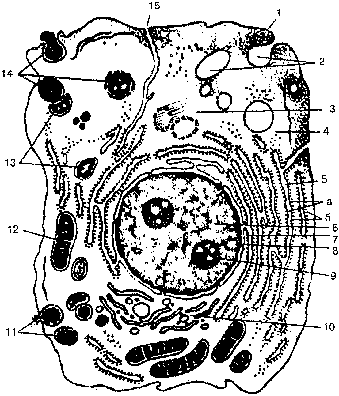
Rice. 2. Scheme of ultramicroscopic cell structure
(according to M.R. Sapin, G.L. Bilich, 1989):
1 - cytolemma (plasma membrane); 2 - pinocytotic vesicles; 3 - centrosome (cellular center, cytocenter); 4 - hyaloplasm; 5 - endoplasmic reticulum (o - endoplasmic reticulum membranes, b - ribosomes); 6- core; 7- connection of the perinuclear space with the cavities of the endoplasmic reticulum; 8 - nuclear pores; 9 - nucleolus; 10 - intracellular mesh apparatus (Golgi complex); 77-^ secretory vacuoles; 12- mitochondria; 7J - lysosomes; 74-three successive stages of phagocytosis; 75 - connection of the cell membrane (cytolemma) with the membranes of the endoplasmic reticulum
Core surrounds cytoplasm, which includes hyaloplasm, organelles and inclusions.
Hyaloplasma- this is the main substance of the cytoplasm, it participates in the metabolic processes of the cell, contains proteins, polysaccharides, nucleic acid, etc.
The permanent parts of the cell that have a specific structure and perform biochemical functions are called organelles. These include the cell center, mitochondria, Golgi complex, endoplasmic (cytoplasmic) reticulum.
Cell center usually located near the nucleus or Golgi complex, it consists of two dense formations - centrioles, which are part of the spindle of a moving cell and form cilia and flagella.
Mitochondria They have the form of grains, threads, sticks, and are formed from two membranes - internal and external. The length of the mitochondrion ranges from 1 to 15 µm, the diameter - from 0.2 to 1.0 µm. The inner membrane forms folds (cristae) in which enzymes are located. The breakdown of glucose, amino acids, and oxidation occurs in mitochondria fatty acids, the formation of ATP (adenosine triphosphoric acid) - the main energy material.
Golgi complex (intracellular reticular apparatus) has the form of bubbles, plates, tubes located around the nucleus. Its function is to transport substances, process them chemically and remove waste products from the cell outside the cell.
Endoplasmic (cytoplasmic) reticulum formed from an agranular (smooth) and granular (granular) network. The agranular endoplasmic reticulum is formed mainly by small cisternae and tubules with a diameter of 50-100 nm, which are involved in the exchange of lipids and polysaccharides. The granular endoplasmic reticulum consists of plates, tubes, cisterns, the walls of which are adjacent to small formations - ribosomes that synthesize proteins.
Cytoplasm also has permanent accumulations of individual substances, which are called cytoplasmic inclusions and are of protein, fat and pigment nature.
The cell, as part of a multicellular organism, performs the main functions: assimilation of incoming substances and their breakdown with the formation of energy necessary to maintain the vital functions of the organism. Cells also have irritability (motor reactions) and are able to multiply by division. Cell division can be indirect (mitosis) or reductional (meiosis).
Mitosis- the most common form of cell division. It consists of several stages - prophase, metaphase, anaphase and telophase. Simple (or direct) cell division - amitosis - occurs rarely in cases where the cell is divided into equal or unequal parts. Meiosis - a form of nuclear division in which the number of chromosomes in a fertilized cell is halved and a restructuring of the cell’s gene apparatus is observed. The period from one cell division to another is called its life cycle.
Fabrics
The cell is part of the tissue that makes up the body of humans and animals.
Textile - it is a system of cells and extracellular structures united by a unity of origin, structure and function.
As a result of the interaction of the organism with the external environment, which developed during the process of evolution, four types of tissues with certain functional features: epithelial, connective, muscular and nervous.
Each organ consists of different tissues that are closely interconnected. For example, the stomach, intestines, and other organs consist of epithelial, connective, smooth muscle and nervous tissues.
Connective tissue of many organs forms stroma, and epithelial - parenchyma. Function digestive system cannot be performed completely if its muscle activity is impaired.
Thus, the various tissues that make up a particular organ ensure that the main function of this organ is performed.